Review
. 2014 Mar 26;4(6):660-77.
doi: 10.7150/thno.8698. eCollection 2014.

Nanotheranostics - application and further development of nanomedicine strategies for advanced theranostics

Affiliations
Review

Nanotheranostics - application and further development of nanomedicine strategies for advanced theranostics

Madaswamy S Muthu et al. Theranostics. .

Abstract

Nanotheranostics is to apply and further develop nanomedicine strategies for advanced theranostics. This review summarizes the various nanocarriers developed so far in the literature for nanotheranostics, which include polymer conjugations, dendrimers, micelles, liposomes, metal and inorganic nanoparticles, carbon nanotubes, and nanoparticles of biodegradable polymers for sustained, controlled and targeted co-delivery of diagnostic and therapeutic agents for better theranostic effects with fewer side effects. The theranostic nanomedicine can achieve systemic circulation, evade host defenses and deliver the drug and diagnostic agents at the targeted site to diagnose and treat the disease at cellular and molecular level. The therapeutic and diagnostic agents are formulated in nanomedicine as a single theranostic platform, which can then be further conjugated to biological ligand for targeting. Nanotheranostics can also promote stimuli-responsive release, synergetic and combinatory therapy, siRNA co-delivery, multimodality therapies, oral delivery, delivery across the blood-brain barrier as well as escape from intracellular autophagy. The fruition of nanotheranostics will be able to provide personalized therapy with bright prognosis, which makes even the fatal diseases curable or at least treatable at the earliest stage.

Keywords: Cancer nanotechnology; Drug targeting; Molecular biomaterials; Molecular imaging; Oral chemotherapy; Pharmaceutical nanotechnology..


Conflict of interest statement

Competing Interests: The authors have declared that no competing interest exists.

Figures

Figure 1
Figure 1
Plasma concentration-time profiles of docetaxel after oral administration to Sprague-Dawley rats at 10 mg/kg dose formulated in the PLGA nanoparticles, PLA-TPGS nanoparticles (NPs), PLGA/montmorillonite NPs, PLA-TPGS/montmorillonite NPs or Taxotere®, compared with the i.v. administration of Taxotere® (n = 5). The therapeutic window is defined as the drug concentration in plasma between the maximum tolerated level (2700 ng/ml) and the minimum effective level (35 ng/ml). Reproduced with permission from Figure 4 of ref. Elsevier Ltd, © (2013).
Figure 2
Figure 2
In vivo pharmacokinetics profiles of docetaxel plasma concentration vs. time after i.v. administration of Taxotere® and the TPGS-emulsified PLA-TPGS NPs formulation using Sprague-Dawley rats at the same docetaxel dose of 10 mg/kg (n = 5). Reproduced with permission from Figure 5 of ref. Bentham Science Publishers Ltd, © (2010).
Figure 3
Figure 3
Schematic diagram of theranostic A) polymer-drug conjugate; B) polymeric nanoparticle; C) solid lipid nanoparticle; D) dendrimer; E) liposome; F) micelle; G) gold nanoparticle; and H) carbon nanotube
Figure 4
Figure 4
Schematic representation of the degradation pathway of PLGA-based nanoparticles in cancer cell. Reproduced with permission from Figure 8 of ref. Elsevier Ltd, © (2014).
Figure 5
Figure 5
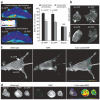
Multimodal imaging of polymeric nanoparticles A) Confocal images of MCF-7 cells treated with the quantum dots and iron oxides loaded PLA-TPGS nanoparticle in vitro (scale bar = 10 μm). A: Bright field image of cells. B: Blue coded stained nuclei. C: Red coded quantum dots from nanoparticles in cytoplasm. D: Complete overlapped image. B) Axial MRI image sections of the MCF-7 grafted tumor bearing mice. Images A and B show the part of the tumor (shown by the arrow) before and after 6 h of administration of the quantum dots and iron oxides-loaded PLA-TPGS nanoparticle into the mice. Images C and D show the kidney (K) and liver (L) part of the mice before and 6 h after the administration of the PLA-TPGS nanoparticle formulation of quantum dots and iron oxides (dosage: 1.5 mg of Cd/kg of body weight or equivalent of 6.0 mg of Fe/kg body weight). The decrease in intensity in the regions of the tumor and liver can be noticed in comparison with the color scale aside. C) Fluorescent images of the various organs. Upper row: control. Lower row: Organs of the mouse treated with the quantum dots and iron oxides-loaded PLA-TPGS nanoparticle (dosage: 1.5 mg of Cd/kg of body weight or equivalent of 6.0 mg of Fe/kg body weight). Reproduced with permission from Figure 2, 5, 6 of ref. Elsevier Ltd, © (2011).
Figure 6
Figure 6
Theranostic approach using dextran coated iron oxide nanoparticles (a) In vivo MRI was performed on mice bearing bilateral green fluorescent protein (9L-GFP) and red fluorescent protein (9L-RFP) tumors before and 24 h after nanoparticles administration. After injection of the probe, there was a significant drop in T2 relaxivity associated with the tumors. Note that T2 relaxation times of muscle tissue remained unchanged. (b) Ex vivo high-resolution MRIs of excised tumors (78 m isotropic). Distinct foci of signal loss (arrows), reflecting probe accumulation, were easily identifiable in tumors derived from mice injected with the probe but not from saline-injected controls. (c) In vivo NIR optical imaging of the same mice as in a produced a high-intensity NIR signal associated with the tumors. This confirmed the delivery of the nanoparticles probe to these tissues. (d) Ex vivo NIR optical imaging demonstrated a significantly higher fluorescence in tumors than in muscle tissue (P = 0.0058). Reproduced with permission from Figure 3 of ref. , Macmilan Publishers Ltd: Nature Medicine, © (2007).
Figure 7
Figure 7
In vitro theranostic delivery using TPGS liposomes A) Field-emission transmission electron microscopy image of (A) an individual quantum dots loaded-TPGS coated liposome in 100 nm scale, and (B) multiple quantum dots-loaded TPGS coated liposomes after the storage in 500 nm scale. B) Confocal laser scanning microscopy images using MCF-7 cells after 2 h incubation with the non-targeted TPGS based multi-functional liposomes (left column) and targeted TPGS based multi-functional liposomes (right column). Row (A): Quantum dots showing the red fluorescence from liposomes distributed in cytoplasm, (B): Channels showing the blue fluorescence from dye stained nuclei, and (C): Merged channels of quantum dots and blue dye. Scale bar = 10 μm. Reproduced with permission from Figure 3, 6 of ref. Elsevier Ltd, © (2012).
Figure 8
Figure 8
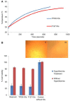
In vitro effect of theranostic micelles loaded with iron oxide nanoparticles (A) Hyperthermia study shows that the time-dependent temperature rise of 1 mg/ml of the iron oxide nanoparticles (IOs)-loaded TPGS and F127 micelles on exposure to 89 kA/m alternating current field at 240 kHz frequency. The specific absorption rate value for the IOs-loaded TPGS and F127 micelles was found to be 51.4 and 25.5 Watt/g. (B) The cytotoxic assay performed after hyperthermia treatment of MCF-7 cancer cells incubated with the IOs-loaded TPGS and F127 micelle and Resovist®. Hyperthermia treatment in all cases leads to significant cell death. Inset image (i) shows the cells incubated with the IOs-loaded TPGS micelles but no AC field applied and inset image (ii) shows the cells incubated with the IOs-loaded TPGS micelles and hyperthermia treatment using AC field. From (ii), it can be seen that after hyperthermia treatment, the cell loses viability and do not attach. Reproduced with permission from Figure 6 of ref. Elsevier Ltd, © (2011).
Figure 9
Figure 9
In vivo imaging of theranostic micelles loaded with iron oxide nanoparticles A) Graph showing the change of T2 values measured from MCF-7 xenograft tumor at the time point before and 45 min, 4 h and 24 h after injection of the iron oxide nanoparticles (IOs)-loaded TPGS and F127 micelle and Resovist®, respectively at a concentration of 5 mg of Fe/Kg of animal weight. It can be found that the T2 value decreases due to accumulation of IOs at tumor by ∼4.3%, 7.3% and 6.9% for Resovist®, the IOs-loaded TPGS and F127 micelles respectively at 45 min after injection. B) T2 mapped images showing the MCF-7 xenograft tumor at the designated time point before and 45 min, 4 h and 24 h after injection of Resovists®, the IOs-loaded TPGS and F127 micelles respectively at a concentration of 5 mg of Fe/Kg of animal weight. The change in T2 in the tumor (marked by the arrow) can be noticed in comparison with the T2 color scale aside. Reprinted with permission from Figure 9, 10 of ref. Elsevier Ltd, © (2011).

Similar articles

Cited by

References

    1. Sumer B, Gao J. Theranostic nanomedicine for cancer. Nanomedicine (Lond) 2008;3(2):137–140. - PubMed
    1. Deveza L, Choi J, Yang F. Therapeutic angiogenesis for treating cardiovascular diseases. Theranostics. 2012;2(8):801–814. - PMC - PubMed
    1. Janib SM, Moses AS, MacKay JA. Imaging and drug delivery using theranostic nanoparticles. Adv Drug Deliv Rev. 2010;62(11):1052–1063. - PMC - PubMed
    1. Yu MK, Park J, Jon S. Targeting strategies for multifunctional nanoparticles in cancer imaging and therapy. Theranostics. 2012;2(1):3–44. - PMC - PubMed
    1. Xie J, Lee S, Chen X. Nanoparticle-based theranostic agents. Adv Drug Deliv Rev. 2010;62(11):1064–79. - PMC - PubMed

Publication types