Review
. 2014 Sep;26(9):557-72.
doi: 10.1111/jne.12157.

Novel aspects of glucocorticoid actions

Affiliations
Review

Novel aspects of glucocorticoid actions

E T Uchoa et al. J Neuroendocrinol. 2014 Sep.

Abstract

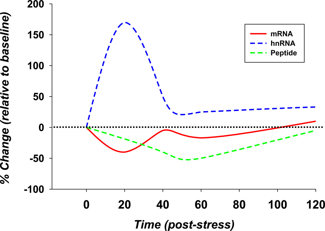
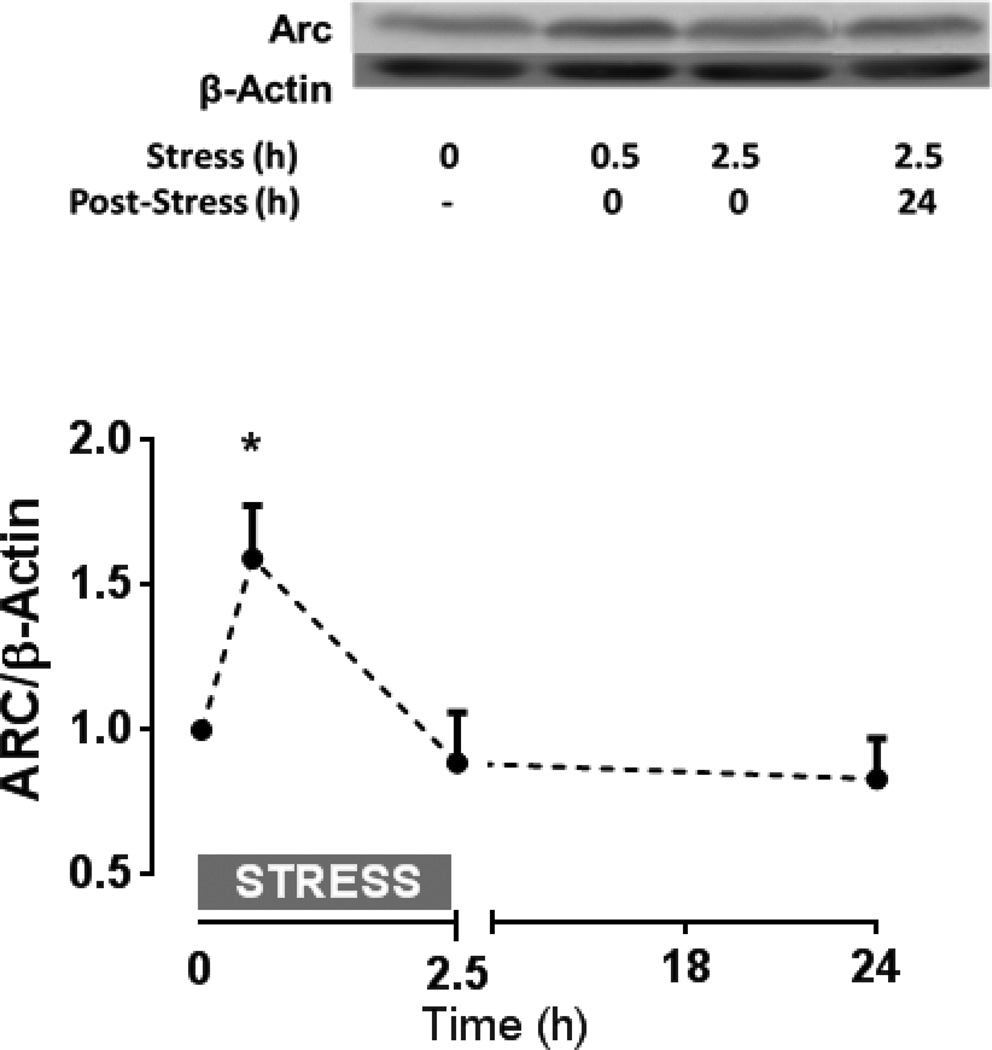
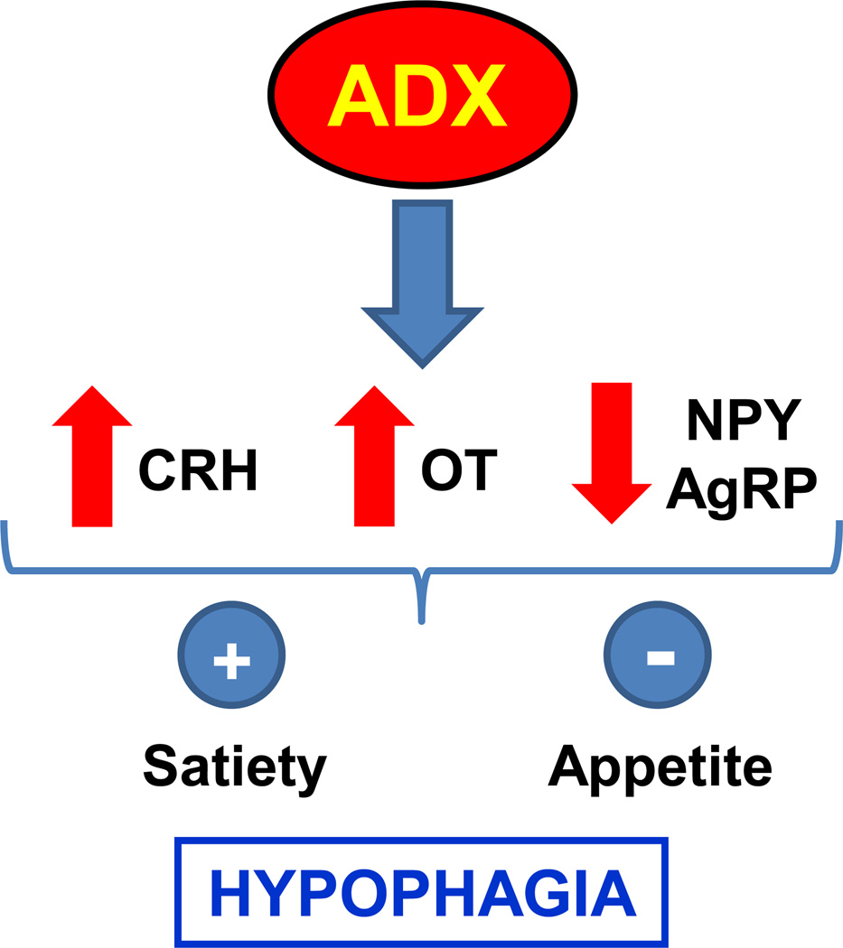
Normal hypothalamic-pituitary-adrenal (HPA) axis activity leading to the rhythmic and episodic release of adrenal glucocorticoids (GCs) is essential for body homeostasis and survival during stress. Acting through specific intracellular receptors in the brain and periphery, GCs regulate behaviour, as well as metabolic, cardiovascular, immune and neuroendocrine activities. By contrast to chronic elevated levels, circadian and acute stress-induced increases in GCs are necessary for hippocampal neuronal survival and memory acquisition and consolidation, as a result of the inhibition of apoptosis, the facilitation of glutamatergic neurotransmission and the formation of excitatory synapses, and the induction of immediate early genes and dendritic spine formation. In addition to metabolic actions leading to increased energy availability, GCs have profound effects on feeding behaviour, mainly via the modulation of orexigenic and anorixegenic neuropeptides. Evidence is also emerging that, in addition to the recognised immune suppressive actions of GCs by counteracting adrenergic pro-inflammatory actions, circadian elevations have priming effects in the immune system, potentiating acute defensive responses. In addition, negative-feedback by GCs involves multiple mechanisms leading to limited HPA axis activation and prevention of the deleterious effects of excessive GC production. Adequate GC secretion to meet body demands is tightly regulated by a complex neural circuitry controlling hypothalamic corticotrophin-releasing hormone (CRH) and vasopressin secretion, which are the main regulators of pituitary adrenocorticotrophic hormone (ACTH). Rapid feedback mechanisms, likely involving nongenomic actions of GCs, mediate the immediate inhibition of hypothalamic CRH and ACTH secretion, whereas intermediate and delayed mechanisms mediated by genomic actions involve the modulation of limbic circuitry and peripheral metabolic messengers. Consistent with their key adaptive roles, HPA axis components are evolutionarily conserved, being present in the earliest vertebrates. An understanding of these basic mechanisms may lead to novel approaches for the development of diagnostic and therapeutic tools for disorders related to stress and alterations of GC secretion.

Keywords: HPA axis; feedback; food intake; glucocorticoids; immune system; neuroplasticity.


Figures

Figure 1
Figure 1
Effect of corticosterone on CRH-stimulated POMC hnRNA in primary cultures of rat anterior pituitary cells (A), and forskolin-stimulated CRH hnRNA in primary cultures of hypothalamic neurones (B). Three-day cultured trypsin-dispersed anterior pituitary cells, maintained in stripped serum for 36h were exposed to 100 nM corticosterone for 18 or 30min before addition of CRH 30 pM for an additional 30 min. Bars represent the mean and SE of POMC hnRNA levels determined by qRT-PCR in 3 cell preparations. In panel B, 10-day cultured fetal rat hypothalamic neuronal cultures were exposed to 100 nM corticosterone for 18h or 30 min before addition of forskolin (Fsk) for an additional 45 min before RNA preparation. Data points are the mean and SE of CRH hnRNA levels, normalized to GAPDH mRNA in 4 experiments. ***, p<0.001 compared with basal; # p< 0.05 lower than Fsk at 0min after log transformation of the data. &, p<0.001 vs CRH at time 0. The horizontal dashed lined represent the SE of maximal stimulated values in the absence of corticosterone.
Figure 2
Figure 2
Time course of the changes in CRH hnRNA (A) and vasopressin (VP) hnRNA (B) after injection of corticosterone (2.8 mg/100 g BW, ip) or vehicle in 48-h adrenalectomised (ADX) or sham operated rats. Note that vehicle injection caused marked increases in CRH hnRNA in ADX but not in intact rats. Data points are the mean and SE of the optical density values obtained from in situ hybridization film autoradiograms in six rats per experimental group. *p<0.01 vs sham; **, p<0.001 vs time 0 and sham; ##, p< 0.01 lower than ADX vehicle. (From Ma and Aguilera, 1999).
Figure 3
Figure 3
Effect of restraint stress on plasma corticosterone, CRH hnRNA levels in the PVN (B), and glucocorticoid receptor recruitment by the CRH and Per 1 promoters in intact male rats. Restraint stress caused marked increases in plasma corticosterone (A), associated with transient increases in CRH hnRNA in the PVN (B). CRH hnRNA was measured by in situ hybridization expressed as optical density (OD) of film autoradiographs (representative images are displayed at the top of data points showing the pooled values in 6 rats). Chromatin immunoprecipitation (ChIP) of microdissected hypothalamic PVN region using anti-GR antibody (IP GR) shows no association of GR with the CRH promoter (solid line and circles), but marked immunoprecipitation of Per 1 promoter (dashed lines open circles) by GR antibodies (C). Immunoprecipitation with phopho-CREB antibody (IP pCREB) yielded high CRH but not Per1 promoter (D). Measurements were performed in basal conditions (time 0), 0.5 and 1h during stress. Data points are the mean and SE of the results of 3 experiments (using pooled hypothalamic tissue from 3 rats per experimental group). The dashed lines correspond to the Per1 promoter, and solid lines show different regions of the CRH promoter. The restraint stress period is shown by the horizontal boxes above the x axis. ***, p< 0.001 vs respective basal; **, p<0.01 vs respective basal; *, p< 0.05 vs. respective basal.
Figure 4
Figure 4
Proposed ‘non-traditional’ glucocorticoid feedback mechanisms. Mechanism 1: Glucocorticoids (GCs) act via non-genomic mechanisms to inhibit PVN CRH neurones, acting via membrane glucocorticoid receptors (pentagon) to mobilize endocannabinoids (ECs), which bind to CB1 receptors and inhibit presaynaptic glutamate release. Mechanism 2: Glucocorticoids act to destabilize preproglucagon mRNA, reducing the magnitude of glucacon-like peptide 1 (GLP-1) excitation of CRH neurones. Mechanism 3: Glucocorticoids act in the periphery (possibly at adipocytes) to generate inhibitory messengers, such as free fatty acids, that can inhibit HPA axis activation secondarily.
Figure 5
Figure 5
Schematic comparing the time course of PPG mRNA degradation (PPG mRNA) with PPG transcription (PPG mRNA) and loss of PVN GLP-1 immunoreactive terminals (peptide). Note that transcriptional effects do not correspond with loss of mRNA, suggesting the mRNA and peptide loss are likely linked to mRNA degradation or turnover.
Figure 6
Figure 6
Effect of acute restraint stress on the Arc (activity-regulated cytoskeleton-associated protein) protein levels in the hippocampus of rats. Acute restraint stress induces an increase of Arc protein levels in rat hippocampus. Sprague Dawley male rats were restrained during 0.5 h or 2.5 h and immediately sacrificed or restrained during 2.5 h and sacrificed 24 h after the restraint. A) Representative immunoblots of homogenates from the hippocampus of stressed rats using anti-Arc and β-actin antibodies. B) Graph shows the relative ratio of Arc levels relative to β-actin. Data represent mean ± SD of n=4 per experimental condition. *p<0.05 when comparing to non-stressed animal (i.e. time= 0).
Figure 7
Figure 7
Schematic illustration showing the hypophagic effect in response to adrenalectomy (ADX) through the stimulation of appetite and the inhibition of satiety pathways, mediated by the increase on the expression of the anorexigenic neuropeptides corticotrophin releasing hormone (CRH) and oxytocin (OT) and the decrease of the orexigenic neuropeptides neuropeptide Y (NPY) and agouti related protein (AgRP).
Figure 8
Figure 8
Schematic illustration describing the interaction between corticosteroids and cytokine expression. Stress exposure directly induces the release of HPA axis hormones and increases expression of cytokines. Although there is emerging evidence to suggest that corticosteroids may enhance cytokine expression under certain circumstances, a primary effect of corticosteroids is suppression of cytokine expression. Cytokines, on the other hand, directly stimulates activation of the HPA axis via actions both intrinsic and extrinsic to the axis, and appear to augment HPA axis sensitivity to later stress challenges.

Similar articles

Cited by

References

    1. Johnson EO, Kamilaris TC, Chrousos GP, Gold PW. Mechanisms of stress: a dynamic overview of hormonal and behavioral homeostasis. Neurosci Biobehav Rev. 1992;16(2):115–130. - PubMed
    1. Smith SM, Vale WW. The role of the hypothalamic-pituitary-adrenal axis in neuroendocrine responses to stress. Dialogues Clin Neurosci. 2006;8(4):383–395. - PMC - PubMed
    1. Lightman SL, Wiles CC, Atkinson HC, Henley DE, Russell GM, Leendertz JA, McKenna MA, Spiga F, Wood SA, Conway-Campbell BL. The significance of glucocorticoid pulsatility. Eur J Pharmacol. 2008;583(2–3):255–262. - PubMed
    1. Joëls M, de Kloet ER. Mineralocorticoid and glucocorticoid receptors in the brain. Implications for ion permeability and transmitter systems. Prog Neurobiol. 1994;43(1):1–36. - PubMed
    1. Hill MN, Tasker JG. Endocannabinoid signaling, glucocorticoid-mediated negative feedback, and regulation of the hypothalamic-pituitary-adrenal axis. Neuroscience. 2012;204:5–16. - PMC - PubMed

Publication types

MeSH terms