. 2015 Jan;25(1):24-32.
doi: 10.1111/bpa.12149. Epub 2014 May 19.

Comorbid rat model of ischemia and β-amyloid toxicity: striatal and cortical degeneration

Affiliations

Comorbid rat model of ischemia and β-amyloid toxicity: striatal and cortical degeneration

Zareen Amtul et al. Brain Pathol. 2015 Jan.

Abstract

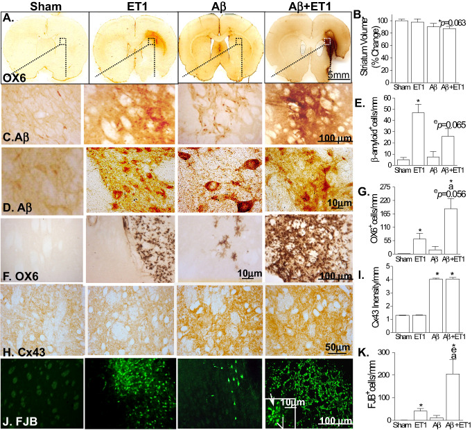
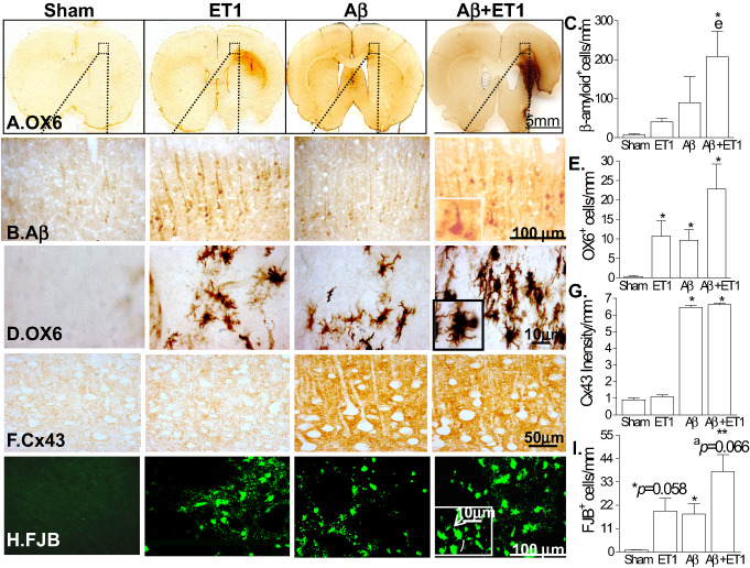
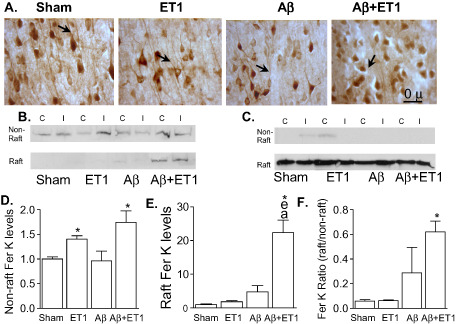
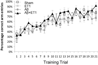
Levels of cerebral amyloid, presumably β-amyloid (Abeta), toxicity and the incidence of cortical and subcortical ischemia increases with age. However, little is known about the severe pathological condition and dementia that occur as a result of the comorbid occurrence of this vascular risk factor and Abeta toxicity. Clinical studies have indicated that small ischemic lesions in the striatum are particularly important in generating dementia in combination with minor amyloid lesions. These cognitive deficits are highly likely to be caused by changes in the cortex. In this study, we examined the viability and morphological changes in microglial and neuronal cells, gap junction proteins (connexin43) and neuritic/axonal retraction (Fer Kinase) in the striatum and cerebral cortex using a comorbid rat model of striatal injections of endothelin-1 (ET1) and Abeta toxicity. The results demonstrated ventricular enlargement, striatal atrophy, substantial increases in β-amyloid, ramified microglia and increases in neuritic retraction in the combined models of stroke and Abeta toxicity. Changes in connexin43 occurred equally in both groups of Abeta-treated rats, with and without focal ischemia. Although previous behavioral tests demonstrated impairment in memory and learning, the visual discrimination radial maze task did not show significant difference, suggesting the cognitive impairment in these models is not related to damage to the dorsolateral striatum. These results suggest an insight into the relationship between cortical/striatal atrophy, pathology and functional impairment.

Keywords: axonal retraction; beta-amyloid; cellular degeneration; infarct; microgliosis.


Figures

Figure 1
Figure 1
Striatal atrophy and pathology: representative coronal sections stained with OX6 at the levels of motor cortex show ventricular enlargement, striatal shrinkage and infarct volume in Aβ+ET1 rats compared with sham rats with normal brain morphology (A). High‐resolution immunostaining images indicate cells demonstrating expression of APP fragments including β‐amyloid (C,D), ramified microglia (OX‐6; F), connexin43 (Cx43; H) and degeneration (FJB; J) in the ipsilateral striatum of Aβ+ET1 rats. Inset in F showed high‐resolution FJB positive cells that appeared to be degenerating neurons (line) and microglia (arrow). Plots demonstrate a quantitative analysis of reduction in the volume of ipsilateral striatum (B), β‐amyloid (E), OX6 (G), Cx43 (I) and FJB (K) immunostaining in the ipsilateral striatum across the groups, *,e,a P < 0.05, (*compared with sham, ecompared with ET1, acompared with Aβ).
Figure 2
Figure 2
Cortical pathology: the low‐magnification images of rat brain sections stained with OX6 in A illustrate the location of the high‐magnification photomicrographs in B–E. Immunostaining indicates cells demonstrating expression of APP fragments (including β‐amyloid; B), ramified microglia (OX‐6; C), connexin43 (Cx43; D) and degeneration (FJB; E) in the ipsilateral neocortex of Aβ+ET1 rats. Insets showed high‐resolution images of β‐amyloid expressing neuronal (B), ramified microglial (C) and FJB positive cells (E) that appeared to be degenerating neurons (E; line) and microglia (E; arrow). Plots in F–I show quantitative analysis of immunostaining in the ipsilateral cortex across the groups, *P < 0.05, **P < 0.01, (*compared with sham, ecompared with ET1, acompared with Aβ).
Figure 3
Figure 3
Axonal retraction: immunostaining of neurons (NeuN) appears to show pyknosis and empty space around shrunken cell bodies and a decrease in the length of neuronal processes (arrows) in the layer V of ipsilateral cortex of Aβ+ET1 rats (A). Western blots show levels of Fer kinase (Fer K) (B) in non‐raft and raft fractions, and flotillin‐1 (C) protein in the raft section of the contralateral (C) and ipsilateral (I) cortices. Plots show quantitative analysis of sham normalized increase in Fer K levels into the ipsilateral non‐raft (D) and raft (E) containing fractions, as well as the ratio of Fer K levels within the raft to non‐raft domains (F) of Aβ+ET1 rats, *P < 0.05, (*compared with sham, ecompared with ET1, acompared with Aβ).
Figure 4
Figure 4
Cognitive impairment: cognitive testing using visual discrimination radial maze task indicates percentage of correct arm entries by sham, ET1, Αβ and Aβ+ET1 rats over the course of 21 training days. Plot shows no differences between any groups. Abbreviations: Alzheimer's disease (AD), amyloid precursor protein (APP), β‐amyloid (Aβ), intracerebroventricular (ICV), endothelin‐1 ( ET1), phosphate‐buffered saline (PBS), avidin–biotin complex (ABC), connexin43 (Cx43), 3,3′‐diaminobenzidine tetrahydrochloride (DAB), fluorojade B (FJB), contralateral (C), ipsilateral (I).

References

    1. Abe K, Tanzi RE, Kogure K (1991) Selective induction of Kunitz‐type protease inhibitor domain‐containing amyloid precursor protein mRNA after persistent focal ischemia in rat cerebral cortex. Neurosci Lett 125:172–174. - PubMed
    1. Akama KT, Albanese C, Pestell RG, Van Eldik LJ (1998) Amyloid beta‐peptide stimulates nitric oxide production in astrocytes through an NFkappaB‐dependent mechanism. Proc Natl Acad Sci U S A 95:5795–5800. - PMC - PubMed
    1. Akundi RS, Candelario‐Jalil E, Hess S, Hull M, Lieb K, Gebicke‐Haerter PJ, Fiebich BL (2005) Signal transduction pathways regulating cyclooxygenase‐2 in lipopolysaccharide‐activated primary rat microglia. Glia 51:199–208. - PubMed
    1. Amtul Z, Hepburn JD (2014) Protein markers to cerebrovascular disruption of neurovascular unit: immunohistochemical and imaging approaches. Rev Neurosci doi: 10.1515/revneuro-2013-0041. [Epub ahead of print]. - DOI - PubMed
    1. Amtul Z, Nikolova S, Gao L, Keeley RJ, Bechberger JF, Fisher AL et al (2014) Comorbid Abeta toxicity and stroke: hippocampal atrophy, pathology, and cognitive deficit. Neurobiol Aging 35:1605–1614. - PubMed

Publication types

MeSH terms