Review
. 2014 Apr;12(2):87-99.
doi: 10.1016/j.jtos.2013.10.007. Epub 2013 Dec 31.

The chemokine receptor CCR7 expressed by dendritic cells: a key player in corneal and ocular surface inflammation

Affiliations
Review

The chemokine receptor CCR7 expressed by dendritic cells: a key player in corneal and ocular surface inflammation

Daniel R Saban. Ocul Surf. 2014 Apr.

Abstract

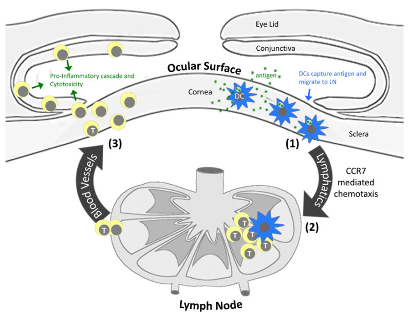
Dendritic cells (DCs) are highly potent stimulators of the immune system, and their contribution as such to the pathogenesis of corneal and ocular surface inflammatory disease has been well established. These vigorous antigen-presenting cells are reliant upon their effective migration from peripheral tissues (e.g., those of the ocular surface) to the lymphoid organs, where immune responses are triggered and can then cause disease. The chemokine receptor CCR7 expressed on DCs has emerged as the master mediator of this highly complex migratory process, and thus it is important in causing corneal and ocular surface inflammation. Furthermore, CCR7 has received considerable attention as a potential therapeutic target, as topically instilled antagonists of this receptor are quite effective therapeutically in a mouse model of ocular allergy. These findings and more are reviewed in the current article. In addition, the understanding regarding CCR7 function in mice and humans, and the biology of DCs that populate the ocular surface are also detailed herein. The involvement of DCs and their expression of CCR7 in corneal and ocular surface diseases such as in ocular allergy, dry eye disease, immune rejection and more, are also reviewed here.

Keywords: CCR7; CD103; T cells; allergic conjunctivitis; conjunctivitis; dendritic cells; dry eye disease; keratitis; ocular allergy; ocular surface.


Conflict of interest statement

Disclosure/Conflict of Interest: Author is inventor on patent application.

Figures

Figure 1
Figure 1
Contribution of DCs and their expression of CCR7 to the pathogenesis of corneal and ocular surface inflammatory disease. DCs that populate the tissues of the ocular surface initiate this pathway via (1) capture of antigen (e.g,, microbial products, allergens, autoantigen, alloantigen, etc.) and subsequent migration to regional lymph nodes via a CCR7 mediated process. (2) Pathogenic T cells are then activated in the LN via migratory DCs and access the ocular surface tissues by way of systemic circulation. (3) Pathogenic T cells, in turn, cause disease by promoting a proinflammatory cascade (e.g., secretion of cytokines, recruitment of myeloid cells, etc.), and causing cytotoxicity of ocular cells.
Figure 2
Figure 2
Antigen-charged DCs express CCR7 to gain access to terminal lymphatic vessels. DCs that populate peripheral tissues, such as the cornea and conjunctiva, will upregulate their expression of CCR7 in inflammation. This allows for chemotactic migration (green arrows) of such DCs toward CCR7 ligands expressed by endothelial cells of terminal lymphatic vessels, and subsequent entrance into the lymphatic system. Terminal lymphatics are not open-ended structures as depicted, and DCs must access them via passing by lymphatic endothelial cells.
Figure 3
Figure 3
CCR7-mediated migration of DCs from peripheral tissues to the LN. (A) General migration path (green arrows) by which DCs migrate from peripheral tissues, e.g., ocular surface, to the LN paracortex. Black dashed box indicates the magnified region of interest. (B) Trafficking of DCs from the afferent lymphatic vessel, to the subcapsular sinus, and into the LN parenchyma. CCR7 ligand gradient indicated in red. Black dashed box indicates paracortical region, where T cells are activated. (C) Final chemotaxis toward the high endothelial venules (HEV). Inside the LN parenchyma, DCs follow a CCR7 ligand gradient toward HEV, where pools of naïve T cells are found. Fibroblastic reticular cells, endothelial cells of the HEV, and DCs themselves all contribute to this CCR7 ligand gradient. Naïve T cells and T regulatory cells also express CCR7, and gain access to the LN via CCR7 ligands found on the luminal side of the HEV.
Figure 4
Figure 4
Ontogeny and function of classical DCs that populate nonlymphoid tissues. Classical DCs that populate interstitial spaces of nonlymphoid tissues are derived from bone marrow precursors, including common DC progenitors (CDP) and/or monocytes. These include CD11b+, CD103+, and CD103+ CD11b+ DCs. This is in contrast to Langerhans cells, with precursors derived from a prenatal lineage.
Figure 5
Figure 5
Classical DCs (CD11c+) that populate the cornea and conjunctiva. (A) Similar to the lung, the tissues of the mouse conjunctiva are populated by CD11b+ and CD103+ DCs. Cells from enzymatically digested tissues are analyzed via flow cytometry. Events are gated on CD11c+ I-A/I-E+ (MHC II), then on CD11c+ autofluorescent-, so that respective CD11b+ and CD103+ DC populations can be visualized. (This figure is adapted from Khandelwal et al doi: 10.1371/journal.pone.0064193.) (B) Characterization of Langerin-expressing DC in the cornea. Langerin-expressing DCs are LCs in the corneal epithelium, whereas ones in the corneal stroma are classical DCs that are likely of bone marrow origin. (This figure is adapted from Hattori et al, doi: 10.1167/iovs.10-6741.)
Figure 6
Figure 6
Working model for the role of DCs in the immunopathogenesis of ocular allergy. DCs play numerous key roles in sensitized individuals. Through migration to the LN via CCR7 (1), DCs activate Th2 cells which, in turn, promote B cell production of IgE antibodies (2) relevant in subsequent immediate hypersensitivity reactions. DC activation of Th2 cells is also important in late phase/chronic responses, as Th2 promote differentiation (3), recruitment and activation of pathogenic eosinophils (4). (This figure is adapted from Saban et al, doi: 10.3109/02713683.2012.747617.)
Figure 7
Figure 7
Schematic depiction of experiments that identified the role of CCR7-mediated migration of DCs on ocular allergy. (A) Exogenous eGFP+ DCs injected subconjunctivally into sensitized wild type mice capture allergen (red) from the ocular surface and express CCR7 (1) to migrate to the LN (2). Such migratory DCs are distinct from LN resident DCs (depicted in gray). Migratory DCs stimulate pathogenic T cells (3) which, in turn, cause disease. (B) Exogenous CCR7 knockout (-/-) DCs (green) injected subconjunctivally into sensitized wild type mice capture allergen (red) from the ocular surface. However, these DCs cannot express CCR7, and their migration to LN is thus inhibited. Activation of pathogenic T cells and consequent clinical disease is also inhibited. This schematic is a depiction of experiments performed by Schlereth et al, doi: 10.1016/j.ajpath.2012.02.015.
Figure 8
Figure 8
CD11b+, but not CD103+, DCs are the dominant in triggering pathogenic T cells involved in ocular allergy. Exogenous CD11b+ or CD103+ DCs (or no DCS) were injected subconjunctivally in sensitized wild type mice. Host mice were challenged with allergen daily for 7 days, and clinical signs were scored at 20 minutes, and 6 and 24 hr post-challenge. (Taken from Khandelwal et al, Khandelwal et al doi: 10.1371/journal.pone.0064193.)
Figure 9
Figure 9
Topically instilled CCR7 antagonist has a robust therapeutic effect against late-phase responses in ocular allergy. Sensitized mice were challenged daily for 4 days. Mice were treated topically with either CCR7 antagonists or an isotype control antibody. Representative pictures on day 3 and day 4 of challenge are shown. Data are taken from Schlereth et al, doi: 10.1016/j.ajpath.2012.02.015.

Similar articles

Cited by

References

    1. Khandelwal P, Blanco-Mezquita T, Emami P, et al. Ocular mucosal CD11b+ and CD103+ mouse dendritic cells under normal conditions and in allergic immune responses. PLoS One. 2013;8:e64193. - PMC - PubMed
    1. Schlereth S, Lee HS, Khandelwal P, Saban DR. Blocking CCR7 at the ocular surface impairs the pathogenic contribution of dendritic cells in allergic conjunctivitis. Am J Pathol. 2012;180:2351–60. - PMC - PubMed
    1. Schaumburg CS, Siemasko KF, De Paiva CS, et al. Ocular surface APCs are necessary for autoreactive T cell-mediated experimental autoimmune lacrimal keratoconjunctivitis. J Immunol. 2011;187:3653–62. - PubMed
    1. Chen Y, Chauhan SK, Soo Lee H, et al. Chronic dry eye disease is principally mediated by effector memory Th17 cells. Mucosal Immunol. 2013 Apr 10; doi: 10.1038/mi.2013.20. - DOI - PMC - PubMed
    1. Stern ME, Schaumburg CS, Pflugfelder SC. Dry eye as a mucosal autoimmune disease. Int Rev Immunol. 2013;32:19–41. - PMC - PubMed

Publication types