. 2014 Jul;27(4):611-20.
doi: 10.1111/pcmr.12250. Epub 2014 May 9.

Epac1 increases migration of endothelial cells and melanoma cells via FGF2-mediated paracrine signaling

Affiliations
Free PMC article

Epac1 increases migration of endothelial cells and melanoma cells via FGF2-mediated paracrine signaling

Erdene Baljinnyam et al. Pigment Cell Melanoma Res. 2014 Jul.
Free PMC article

Abstract

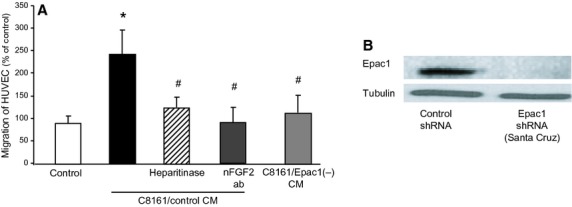
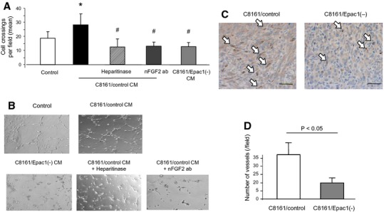
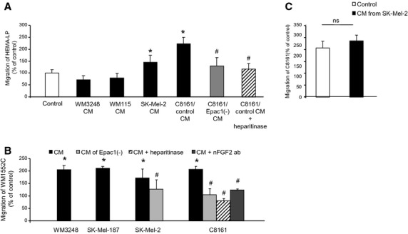
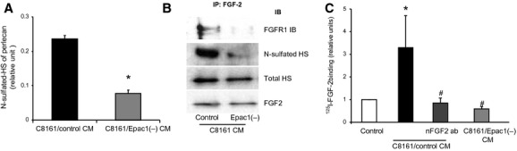
Fibroblast growth factor (FGF2) regulates endothelial and melanoma cell migration. The binding of FGF2 to its receptor requires N-sulfated heparan sulfate (HS) glycosamine. We have previously reported that Epac1, an exchange protein activated by cAMP, increases N-sulfation of HS in melanoma. Therefore, we examined whether Epac1 regulates FGF2-mediated cell-cell communication. Conditioned medium (CM) of melanoma cells with abundant expression of Epac1 increased migration of human umbilical endothelial cells (HUVEC) and melanoma cells with poor expression of Epac1. CM-induced increase in migration was inhibited by antagonizing FGF2, by the removal of HS and by the knockdown of Epac1. In addition, knockdown of Epac1 suppressed the binding of FGF2 to FGF receptor in HUVEC, and in vivo angiogenesis in melanoma. Furthermore, knockdown of Epac1 reduced N-sulfation of HS chains attached to perlecan, a major secreted type of HS proteoglycan that mediates the binding of FGF2 to FGF receptor. These data suggested that Epac1 in melanoma cells regulates melanoma progression via the HS-FGF2-mediated cell-cell communication.

Keywords: Epac; FGF2; angiogenesis; cell-cell communication; heparan sulfate; human umbilical vein endothelial cells; migration; paracrine signaling.


Figures

Figure 1
Figure 1
Epac1 in melanoma cells increases migration of endothelial cells via cell/cell communication. (A) CM of C8161 (C8161/control CM) increased migration of human umbilical vein endothelial cells (HUVEC). Epac1 knockdown in C8161 cells (C8161/Epac1(−) CM) inhibited the CM-induced migration. The CM-induced increase in migration was inhibited by the neutralizing antibody against FGF2 [nFGF2 ab (25 μg/ml)], and heparitinase (0.08 U/ml). *P < 0.05 versus control, #P < 0.05 versus C8161/control CM, n = 4. (B) Western blot of C8161 cells with stable knockdown of Epac1 performed with lentivirus-based shRNA induction.
Figure 2
Figure 2
Epac1 in melanoma cells activates angiogenesis. (A) C8161/control CM increased tube formation of human umbilical vein endothelial cells (HUVEC). C8161/Epac1(−) CM showed reduced tube formation compared to C8161/control CM. The C8161/control CM-induced tube formation was inhibited by nFGF2 ab (25 μg/ml), and by heparitinase (0.08 U/ml). C8161/Epac1(−) CM showed reduced tube formation compared with C8161/control CM.*P < 0.01 versus control medium. #P < 0.01 versus C8161/control CM, n = 4. (B) Representative images of HUVEC tube formation described in A. (C and D) Epac1 knockdown reduces angiogenesis in vivo. C8161 cells with or without Epac1 knockdown (1 × 106 cells) were inoculated in the interscapular region of BALB/c mice. One week after the inoculation, tumor was removed. (C) Immunohistochemical images with anti-CD31 staining for the detection of endothelial cells are shown. White arrows indicate CD31-positive cells stained brown. Scale bar: 100 μm. (D) The number of microvessels in each mouse was counted with the positively stained cells in 10 different fields, n = 4.
Figure 3
Figure 3
Epac1 in melanoma cells increases migration of melanocytes/other melanoma cells. (A) Conditioned media of indicated melanoma cell lines were used for the Boyden chamber migration assay of HEMA-LP cells. Conditioned media from SK-Mel-2 and C8161 cells, but not those from WM3248 and WM115 cells, increased migration of HEMA-LP cells. Knockdown of Epac1 (C8161/Epac1(−) CM) as well as heparitinase inhibited the CM-induced migration. *P < 0.05 versus control medium, #P < 0.05 versus C8161/control CM, n = 4. (B) Conditioned media of indicated melanoma cell lines were used for the Boyden chamber assay of WM1552C cells. Conditioned media of all cell lines examined increased migration of WM1552C cells. Knockdown of Epac1 inhibited migration induced by CM derived from SK-Mel-2 and C8161 cells. Heparitinase and the nFGF-2 antibody suppressed migration induced by CM of C8161 cells.*P < 0.05 versus control medium, #P < 0.05 versus CM, n = 4. (C) The Boyden chamber assay showed that CM of SK-Mel-2 cells did not increase migration of C8161 cells, n = 4.
Figure 4
Figure 4
Epac1 enhances the binding of fibroblast growth factor (FGF2) to FGF receptor via N-sulfation of HS. (A) Perlecan was isolated from the DEAE chromatography fractions using a polyclonal antiperlecan antibody. The presence of HS chains on perlecan was detected using an anti-HS-specific antibody (10E4). Epac1 knockdown reduced the amount of N-sulfated HS attached to perlecan. *P < 0.05 versus C8161/control CM, n = 8. (B) CM of C8161 was subjected to immunoprecipitation with the antibody against FGF2 followed by Western blot for indicated antibodies. Both N-sulfated HS and FGF receptor 1 (FGFR1) attached to FGF2 were reduced by Epac1 knockdown whereas the amount of FGF2 in the CM was not different. (C) The binding assay for FGF2 in human umbilical vein endothelial cells (HUVEC) was performed with indicated CM. C8161/control CM increased the binding of FGF2 to HUVEC. The neutralizing antibody for FGF2 (nFGF2 ab) and knockdown of Epac1 inhibited the CM-induced FGF2 binding. *P < 0.05 versus control medium, #P < 0.05 versus C8161/control CM, n = 4.
Figure 5
Figure 5
Epac1-rich melanoma cells support survival of Epac1-poor melanoma cells. (A) Tumor growth of WM1552C and SK-Mel-2 cells expressing Red Fluorescent Protein (RFP) and Green Fluorescent Protein (GFP) is shown. A mixture of indicated cells was injected in the right dorsolateral flank region in athymic BALB/c nude mice. Tumor size was measured twice a week to calculate tumor volume. Tumor failed to grow in the mixture of RFP- and GFP-labeled WM1552C. (B and C) Representative images of the tumors in the 12 weeks after the inoculation are shown. The mixture of RFP-labeled WM1552C cells and GFP-labeled SK-Mel-2 cells formed a tumor. (D) Representative images of coimmunostaining for RFP and GFP of the tumors formed by the indicated cell mixtures. Blue indicates 6-diamidino-2-phenylindole (DAPI) staining.

References

    1. Aviezer D, Iozzo RV, Noonan DM. Yayon A. Suppression of autocrine and paracrine functions of basic fibroblast growth factor by stable expression of perlecan antisense cDNA. Mol. Cell. Biol. 1997;17:1938–1946. - PMC - PubMed
    1. Baljinnyam E, Iwatsubo K, Kurotani R, Wang X, Ulucan C, Iwatsubo M, Lagunoff D. Ishikawa Y. Epac increases melanoma cell migration by a heparan sulfate-related mechanism. Am. J. Physiol. Cell Physiol. 2009;297:C802–C813. - PMC - PubMed
    1. Baljinnyam E, De Lorenzo MS, Xie LH, Iwatsubo M, Chen S, Goydos JS, Nowycky MC. Iwatsubo K. Exchange protein directly activated by cyclic AMP increases melanoma cell migration by a Ca2 + -dependent mechanism. Cancer Res. 2010;70:5607–5617. - PubMed
    1. Baljinnyam E, Umemura M, De Lorenzo MS, Iwatsubo M, Chen S, Goydos JS. Iwatsubo K. Epac1 promotes melanoma metastasis via modification of heparan sulfate. Pigment Cell Melanoma Res. 2011;24:680–687. - PubMed
    1. Bos JL. Epac proteins: multi-purpose cAMP targets. Trends Biochem. Sci. 2006;31:680–686. - PubMed

Publication types

MeSH terms

Substances