Review
. 2014 Apr 10;157(2):300-311.
doi: 10.1016/j.cell.2014.01.070.

Atomistic autophagy: the structures of cellular self-digestion

Affiliations
Review

Atomistic autophagy: the structures of cellular self-digestion

James H Hurley et al. Cell. .

Abstract

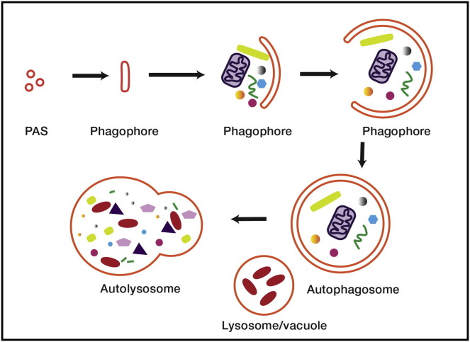
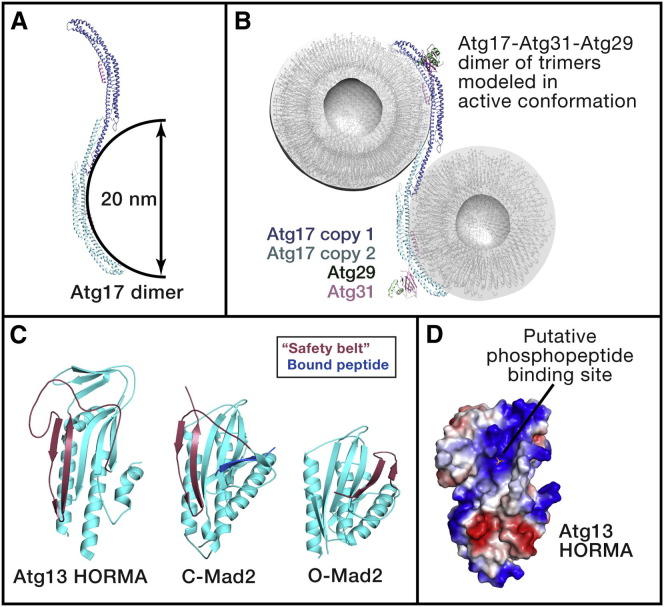
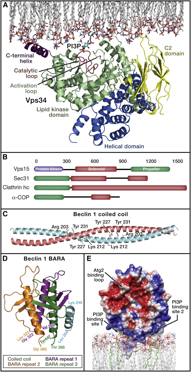
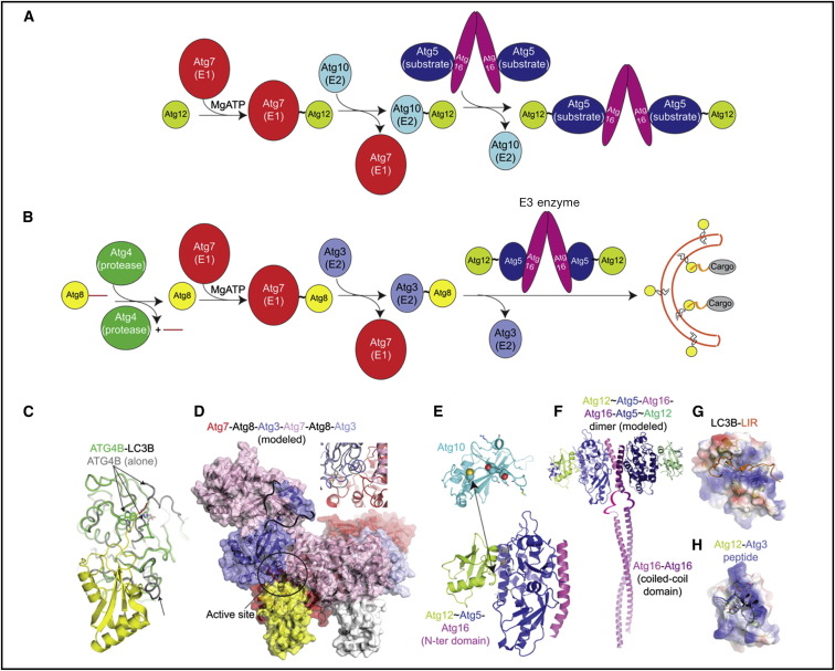
Autophagy is directed by numerous distinct autophagy-related (Atg) proteins. These transmit starvation-induced signals to lipids and regulatory proteins and assemble a double-membrane autophagosome sequestering bulk cytoplasm and/or selected cargos destined for degradation upon autophagosome fusion with a vacuole or lysosome. This Review discusses the structural mechanisms by which Atg proteins sense membrane curvature, mediate a PI(3)P-signaling cascade, and utilize autophagy-specific ubiquitin-like protein cascades to tether proteins to autophagosomal membranes. Recent elucidation of molecular interactions enabling vesicle nucleation, elongation, and cargo recruitment provides insights into how dynamic protein-protein and protein-membrane interactions may dictate size, shape, and contents of autophagosomes.


Figures

Figure 1
Figure 1
Autophagy Autophagy is thought to commence with the clustering of membrane vesicles at the PAS. Membrane fusion leads to the formation of the open double-membrane sheet known as the phagophore. The phagophore expands and matures into the closed autophagosome. Finally, the autophagosome fuses with the lysosome, forming the autolysosome and leading to the degradation of the contents of the autophagosome.
Figure 2
Figure 2
The Atg1 Autophagy Initiation Complex (A) Atg17 is the main scaffold for the Atg1 complex and for the assembly of the PAS for bulk autophagy in yeast. This image shows the complementarity between the shape of the Atg17 dimer (with Atg29 and Atg31 removed for clarity) and a pair of 20 nm diameter vesicles (Ragusa et al., 2012). (B) A model for the putative disinhibited conformation of the Atg17-Atg31-Atg29 complex bound to two 20 nm vesicles. Atg29 and Atg31 were moved from their crystallographic positions, which sterically collide with the docked vesicles, into a conformation where they do not interfere with binding. (C) Structure of the HORMA domains of Atg13 (left) (Jao et al., 2013) and Mad2 (Sironi et al., 2002), with the latter in the closed and open conformations, respectively. The conformationally variable safety belt region is shown in red, and a peptide from Mad1 bound to Mad2 is shown in the central panel in blue. (D) Surface model of the Atg13 HORMA domain (blue is electropositive and red electronegative) and crystallographic sulfate ion as a marker for a putative phosphopeptide-binding site (Jao et al., 2013).
Figure 3
Figure 3
Complexes of PI(3)P Synthesis and Recognition (A) Crystal structure of the catalytic core of Drosophila Vps34, consisting of the catalytic and helical domains (Miller et al., 2010). The C2 domain was not present in the crystal structure and was modeled as described on the basis of the C2 domain of a related PI 3-kinase (Miller et al., 2010). Vps34 is shown docked to the membrane in a conformation in which the C-terminal helix has been moved from its crystallographic position into its putative active conformation as bound to lipids. (B) Most of the structure of the Vps15 subunit is unknown, with the exception of the propeller domain. The schematic shows that Vps15 resembles vesicle coat proteins in its overall domain architecture, with the addition of a protein kinase domain. (C) The antiparallel coiled-coil dimer of beclin 1 (Li et al., 2012). Tyr residues that are phosphorylated by the EGFR are highlighted (Wei et al., 2013), as are basic residues that might potentially form stabilizing cross-dimer interactions with the phospho-Tyr. (D) The pseudo 3-fold symmetric BARA domain of human beclin 1 (Huang et al., 2012). Each of the pseudo 3-fold repeats is colored differently. Four helices are present in this structure because it also includes the most C-terminal portion of the coiled-coil domain. (E) Surface model of K. lactis Hsv2 (Baskaran et al., 2012, Krick et al., 2012), which serves as a structural model for Atg18 and human WIPI proteins. The two PI(3)P-binding sites are highlighted.
Figure 4
Figure 4
Structures, Mechanisms, and Functions of Ubiquitin-like Protein Conjugation Cascades in Autophagy (A) Atg12 ligation pathway progresses first via a thioester-linked Atg7∼Atg12 intermediate and then via a thioester-linked Atg10∼Atg12 intermediate, from which Atg12 is ligated to Atg5, which binds Atg16. (B) Atg8/LC3 ligation pathway involves processing by Atg4, activation by Atg7, conjugation to Atg3, and ligation to phosphatidylethanolamine (PE) facilitated by the Atg12∼Atg5-Atg16 oligomer. Atg8/LC3 (represented in yellow circles) ligation to PE promotes expansion of the phagophore membrane and recruits cargos to autophagosomes. (C) Structural superposition of free ATG4B (gray) and a complex between ATG4B (green) and the precursor form of LC3 (yellow, with C-terminal extension in red). The ATG4 Cys-His-Asp catalytic triad and Trp clamp are shown in sticks (Kumanomidou et al., 2006, Satoo et al., 2009, Sugawara et al., 2005). Arrows highlight regions of ATG4B conformational activation. (D) Structural model for Atg8 (yellow and white) transfer between Atg7 (the two protomers in the homodimer colored red and pink) and Atg3 (two bound per Atg7 homodimer, colored slate and light blue) (Hong et al., 2011, Kaiser et al., 2013, Noda et al., 2011). One of two active sites is circled, from which Atg8 (yellow) is transferred between the active site of Atg7 (red) to Atg3 (slate) bound to the N-terminal domain of the opposite Atg7 in the dimer (pink). Missing Atg3 loops, ribbons. Inset, close-up of active site, superimposed with free Atg3 to highlight conformational activation. Modeling suggests similar transfer of Atg12 to Atg10. (E) Structures of K. marxianus Atg10 (cyan) and human Atg12 (lime)∼Atg5 (blue)-Atg16 (magenta) (Hong et al., 2012, Noda et al., 2013, Otomo et al., 2013, Yamaguchi et al., 2012). Arrow highlights the Atg3 cysteine, from which Atg12 is transferred, to the Atg5 target. Spheres, Atg10’s Cys; positions that can be crosslinked to Atg5; sticks, residues implicated in Atg5 binding (from yeast Atg12, lime; from human and K. marxianus Atg5, gray and white). (F) Model of an Atg12∼Atg5-Atg16 dimer, based on crystal contacts Atg12∼Atg5-Atg16 (N-terminal domain) structures (Metlagel et al., 2013, Otomo et al., 2013), connected by ribbons to Atg16 coiled coil (Fujioka et al., 2010). (G) Surface of LC3B colored by electrostatic potential, bound to LIR motif from p62 (orange) (Ichimura et al., 2008, Pankiv et al., 2007). (H) Surface of Atg12 colored by electrostatic potential, bound to Atg3 (slate) flexible region (Metlagel et al., 2013).

Similar articles

Cited by

References

    1. Baskaran S., Ragusa M.J., Boura E., Hurley J.H. Two-site recognition of phosphatidylinositol 3-phosphate by PROPPINs in autophagy. Mol. Cell. 2012;47:339–348. - PMC - PubMed
    1. Behrends C., Sowa M.E., Gygi S.P., Harper J.W. Network organization of the human autophagy system. Nature. 2010;466:68–76. - PMC - PubMed
    1. Bestebroer J., V’kovski P., Mauthe M., Reggiori F. Hidden behind autophagy: The unconventional role of ATG proteins. Traffic. 2013;14:1029–1041. - PMC - PubMed
    1. Boya P., Reggiori F., Codogno P. Emerging regulation and functions of autophagy. Nat. Cell Biol. 2013;15:713–720. - PMC - PubMed
    1. Brownell J.E., Sintchak M.D., Gavin J.M., Liao H., Bruzzese F.J., Bump N.J., Soucy T.A., Milhollen M.A., Yang X., Burkhardt A.L. Substrate-assisted inhibition of ubiquitin-like protein-activating enzymes: the NEDD8 E1 inhibitor MLN4924 forms a NEDD8-AMP mimetic in situ. Mol. Cell. 2010;37:102–111. - PubMed

Publication types

Substances