. 2014 Apr 10;157(2):433-446.
doi: 10.1016/j.cell.2014.01.067.

Intestinal brush border assembly driven by protocadherin-based intermicrovillar adhesion

Affiliations

Intestinal brush border assembly driven by protocadherin-based intermicrovillar adhesion

Scott W Crawley et al. Cell. .

Abstract

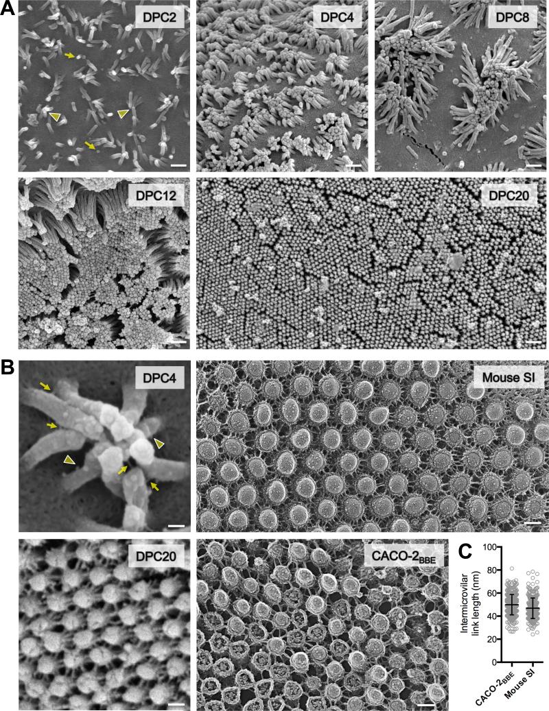
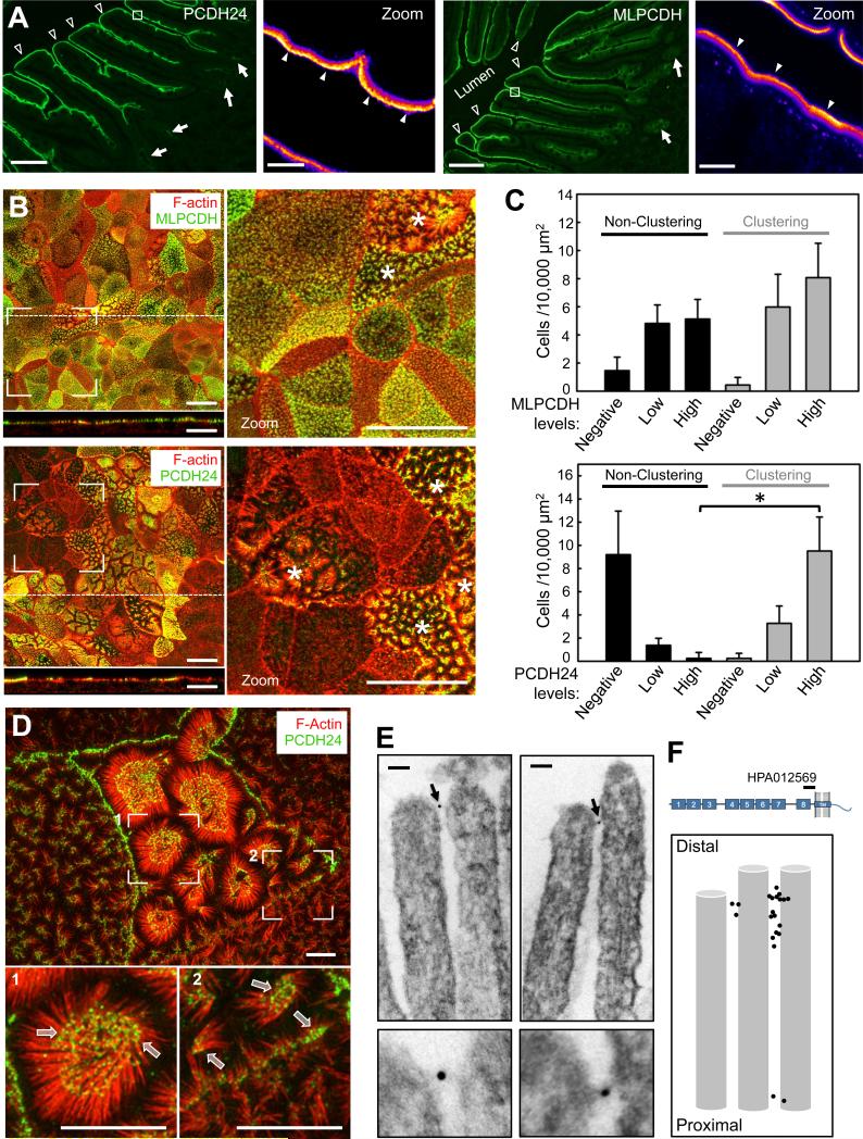
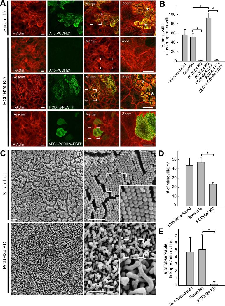
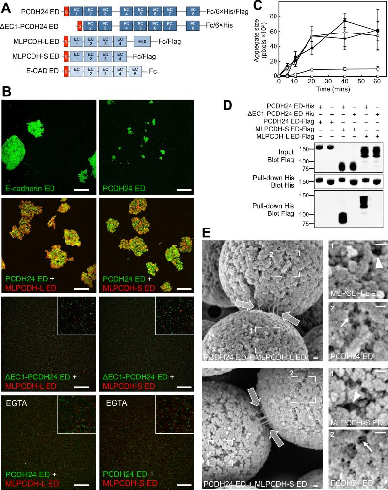
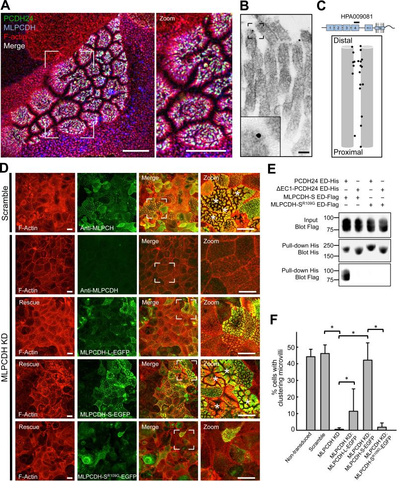
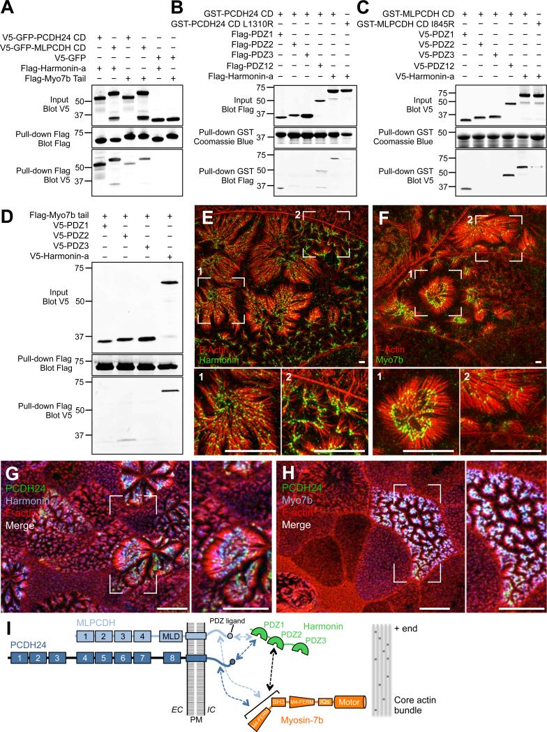
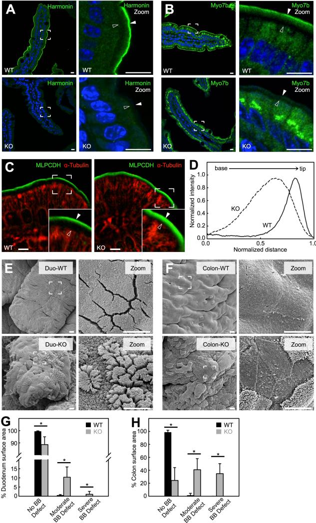
Transporting epithelial cells build apical microvilli to increase membrane surface area and enhance absorptive capacity. The intestinal brush border provides an elaborate example with tightly packed microvilli that function in nutrient absorption and host defense. Although the brush border is essential for physiological homeostasis, its assembly is poorly understood. We found that brush border assembly is driven by the formation of Ca(2+)-dependent adhesion links between adjacent microvilli. Intermicrovillar links are composed of protocadherin-24 and mucin-like protocadherin, which target to microvillar tips and interact to form a trans-heterophilic complex. The cytoplasmic domains of microvillar protocadherins interact with the scaffolding protein, harmonin, and myosin-7b, which promote localization to microvillar tips. Finally, a mouse model of Usher syndrome lacking harmonin exhibits microvillar protocadherin mislocalization and severe defects in brush border morphology. These data reveal an adhesion-based mechanism for brush border assembly and illuminate the basis of intestinal pathology in patients with Usher syndrome. PAPERFLICK:


Figures

Figure 1
Figure 1. Enterocyte BB microvilli cluster during differentiation and are connected by thread-like links
(A) SEM of CACO-2BBE cells from a differentiation time series; days post confluency (DPC) are indicated. Yellow arrows point to initial microvillar membrane buds, arrowheads to distal tip contact of longer microvilli. Scale bars, 500 nm. (B) High magnification images of CACO-2BBE cells and native intestinal tissue. Left upper: SEM of a microvillar cluster from 4-DPC CACO-2BBE cell. Yellow arrows point to intact intermicrovillar adhesion links, arrowheads to unpaired or broken links. Left bottom: SEM of a 20-DPC CACO-2BBE monolayer. Right upper: Freeze-etch EM of the BB from mouse small intestinal tissue. Right lower: Freeze-etch EM of the BB from 20-DPC CACO-2BBE cells. Scale bars, 100 nm. (C) Intermicrovillar link lengths from CACO-2BBE and mouse intestinal tissue freeze-etch EM images (mean ± SD). See also Figure S1.
Figure 2
Figure 2. MLPCDH and PCDH24 localize to the BB in both native intestinal tissue and CACO-2BBE monolayers
(A) Confocal microscopy of human duodenal tissue stained for PCDH24 and MLPCDH. Arrows point to intestinal crypts, open arrowheads to villi, filled arrowheads to the enrichment of PCDH24 and MLPCDH at the distal tips of the microvilli. Boxed regions denote zoomed heat map images of the BB. Scale bars, 100 μm; zoom, 5 μm. (B) Confocal images of 12-DPC cells stained for F-actin and either MLPCDH (top) or PCDH24 (bottom). Dashed lines represent where x-z sections were taken (shown below the en face images). Asterisks denote cells undergoing robust microvillar clustering. Scale bars, 20 μm. (C) Microvillar clustering as a function of endogenous expression levels of MLPCDH (top; n = 995 cells) or PCDH24 (bottom; n = 1146 cells) in 12-DPC monolayers (mean ± SD). *p < 0.0001, t test. (D) Super-resolution microscopy of 12-DPC cells stained for F-actin and PCDH24. Outlined arrows point to distal tip enrichment of PCDH24 in clustering microvilli. Scale bars, 5 μm. (E) Immuno-gold TEM of PCDH24-labeled 12-DPC cells. Bottom panels show zoomed image of highlighted gold particles. Scale bars, 50 nm. (F) Relative positions of gold-labeled particles along the microvillar axis. See also Figure S2.
Figure 3
Figure 3. Stable shRNA knockdown of PCDH24 in CACO-2BBE cells abolishes microvillar clustering and disrupts BB formation
(A) Confocal images of 12-DPC cells stably expressing either a scramble shRNA construct or an shRNA targeting PCDH24. Scramble and PCDH24 KD cells have been stained for F-actin and PCDH24, while PCDH24 KD cell lines expressing rescue constructs have been stained for F-actin only. Asterisks denote cells undergoing robust microvillar clustering. Scale bars, 15 μm (B) Microvillar clustering in non-transduced, scramble, PCDH24 KD, and PCDH24 rescue cell lines. For quantification of rescue lines, only EGFP-positive cells were scored (mean ± SD). Non-transduced cells n = 2639, scramble control cells n = 1019, PCDH24 KD cells n = 1056, PCDH24 KD rescue PCDH24-EGFP n = 115, PCDH24 KD rescue ΔEC1 PCDH24-EGFP cells n = 160; *p values < 0.0001, t test. (C) SEM images of 20-DPC CACO-2BBE cells stably expressing either a scramble shRNA construct (top) or an shRNA targeting PCDH24 (bottom). Scale bars, 1 μm. (D) Microvillar packing density in non-transduced, scramble and PCDH24 KD stably transduced 20-DPC cells (mean ± SD). Area quantified and number of microvilli counted: non-transduced cells area = 150 μm2, microvilli n = 6599; scramble cells area = 125 μm2, microvilli n = 5919; PCDH24 KD cells area = 225 μm2, cells microvilli n = 5318; *p value < 0.0001, t test. (E) Observable links at the distal tips of microvilli in non-transduced, scramble and PCDH24 KD cell lines. Microvilli counted from non-transduced cells n = 100, scramble control cells n = 94 and PCDH24 KD cells n = 110; *p value < 0.0001, t test. See also Figure S3.
Figure 4
Figure 4. PCDH24 and MLPCDH interact to form Ca2+-dependent trans heterophilic adhesion complexes
(A) Constructs used in bead aggregation and pull-down assays. The signal sequences (S) of the cadherin EDs are shown in red. The long isoform of MLPCDH contains a juxtamembrane mucin-like repeat domain (MLD). (B) Confocal images of ED-coated fluorescent bead aggregation assays at the 60 min time point. Insets shown in selected panels are zoomed images to show the presence of individual beads. Scale bars, 250 μm. (C) Bead aggregate size as a function of time. n=three independent experiments. (D) Pull-down analysis using the EDs of PCDH24 and both isoforms of MLPCDH. (E) SEM images of in vitro reconstituted trans heterophilic adhesion complexes between beads coated with the ED of PCDH24 and either MLPCDHL (top) or MLPCDH-S (bottom). White outlined arrows point to adhesion links at bead-bead interfaces. Box 1 (right upper) confirms the identity of the denoted beads as being coated with MLPCDH ED isoforms, given the lack of intra-bead bonds (see arrowheads). Box 2 (right lower) shows PCDH24-coated beads that possess an extensive network of intra-bead bonds (see arrows). Scale bars, 100 nm. See also Figure S4.
Figure 5
Figure 5. MLPCDH colocalizes with PCDH24 and is necessary for microvillar clustering in CACO-2BBE cells
(A) Confocal microscopy of 12-DPC CACO-2BBE monolayers stained for PCDH24, MLPCDH and F-actin. Scale bars, 10 μm. (B) Immuno-gold TEM of MLPCDH-labeled 12-DPC CACO-2BBE cells. Inset shows a zoomed image of a gold particle. Scale bar, 50 nm. (C) Relative positions of gold-labeled particles along the microvillar axis. (D) Confocal images of 12-DPC CACO-2BBE cells stably expressing either a scramble shRNA construct or an shRNA targeting MLPCDH. Scramble and MLPCDH KD cells have been stained for F-actin and MLPCDH, while MLPCDH KD cell lines expressing rescue constructs have been stained for F-actin and GFP. Asterisks denote cells undergoing robust microvillar clustering. Scale bars, 15 μm (E) Pull-down analysis of the MLPCDH-SR109G mutant ED interaction with the PCDH24 ED. (F) Microvillar clustering in non-transduced, scramble, MLPCDH KD, and MLPCDH rescue CACO-2BBE cell lines. For quantification of rescue cell lines, only EGFP-positive cells were scored (mean ± SD). Non-transduced cells n = 1857, scramble control cells n = 963, MLPCDH KD cells n = 1324, MLPCDH KD rescue MLPCDH-L-EGFP n = 441, MLPCDH KD rescue MLPCDH-S-EGFP n = 1141, MLPCDH KD rescue MLPCDH-SR109G-EGFP cells n = 520; *p values < 0.0001, t test. See also Figure S3.
Figure 6
Figure 6. Microvillar protocadherins form a complex with harmonin and Myo7b at the tips of BB microvilli
(A) Pull-down analysis of the interactions of the CDs of PCDH24 and MLPCDH with harmonin-a and the cargo-binding tail of Myo7b. (B and C) Mapping the domains of harmonin-a that interact with the CDs of PCDH24 and MLPCDH. (D) Mapping the domain of harmonin-a that interacts with Myo7b. (E and F) Super-resolution microscopy of 12-DPC CACO-2BBE cells stained for F-actin and harmonin or Myo7b. Scale bars, 3 μm. (G and H) Confocal microscopy of 12-DPC CACO-2BBE cells stained for PCDH24, F-actin, and harmonin or Myo7b. Scale bars, 10 μm. (I) Cartoon summary of interactions between the CDs of microvillar protocadherins, harmonin and Myo7b. See also Figure S6.
Figure 7
Figure 7. Harmonin KO mice exhibit defects in BB morphology
(A and B) Confocal images of villi from WT (top) and harmonin KO (bottom) stained for harmonin, Myo7b and DAPI (blue). Boundaries of the BB are denoted with filled arrowheads (microvillar tips) and open arrowheads (terminal web) in the zoomed images (right panels). Scale bars, 10 μm. (C) Confocal images of villi from WT (left) and harmonin KO (right) littermates stained for MLPCDH and α-tubulin. In zoomed images microvillar tips are marked with filled arrowheads and the terminal web is marked with open arrowheads (insets). Scale bars, 5 μm. (D) Line scans of MLPCDH immunofluorescence intensity within the BB of WT and harmonin KO. (E) SEM images of the apical surface of villi from the duodenum (Duod) of WT (top) and harmonin KO (bottom). (F) SEM of the proximal colon apical surface from WT (top) and harmonin KO (bottom) mice. Scale bars, 5 μm. (G and H) Degree of BB defects exhibited in the duodenum (G) and colon (H) of WT and harmonin KO (mean ± SD). No BB defect was defined as an apical surface that possessed well-packed microvilli of uniform length; moderate BB defect as apical microvilli that were disheveled in appearance; severe BB defect as areas that lacked microvilli. Surface area measured: WT SI = 4,001,746 μm2, Harmonin KO SI = 296,867 μm2, WT colon = 511,465 μm2, Harmonin KO colon = 975,557 μm2; n = 4 animals for each genotype. *p value < 0.0001, t test. See also Figure S7.

References

    1. Alagramam KN, Goodyear RJ, Geng R, Furness DN, van Aken AF, Marcotti W, Kros CJ, Richardson GP. Mutations in protocadherin 15 and cadherin 23 affect tip links and mechanotransduction in mammalian sensory hair cells. PLoS One. 2011;6:e19183. - PMC - PubMed
    1. Bailey DS, Freedman AR, Price SC, Chescoe D, Ciclitira PJ. Early biochemical responses of the small intestine of coeliac patients to wheat gluten. Gut. 1989;30:78–85. - PMC - PubMed
    1. Belyantseva IA, Boger ET, Naz S, Frolenkov GI, Sellers JR, Ahmed ZM, Griffith AJ, Friedman TB. Myosin-XVa is required for tip localization of whirlin and differential elongation of hair-cell stereocilia. Nat Cell Biol. 2005;7:148–156. - PubMed
    1. Berndorff D, Gessner R, Kreft B, Schnoy N, Lajous-Petter AM, Loch N, Reutter W, Hortsch M, Tauber R. Liver-intestine cadherin: molecular cloning and characterization of a novel Ca(2+)-dependent cell adhesion molecule expressed in liver and intestine. J Cell Biol. 1994;125:1353–1369. - PMC - PubMed
    1. Bitner-Glindzicz M, Lindley KJ, Rutland P, Blaydon D, Smith VV, Milla PJ, Hussain K, Furth-Lavi J, Cosgrove KE, Shepherd RM, et al. A recessive contiguous gene deletion causing infantile hyperinsulinism, enteropathy and deafness identifies the Usher type 1C gene. Nat Genet. 2000;26:56–60. - PubMed

Publication types

MeSH terms