Review
. 2014 May;41(2):315-26.
doi: 10.1016/j.ucl.2014.01.007.

Screening and detection advances in magnetic resonance image-guided prostate biopsy

Affiliations
Review

Screening and detection advances in magnetic resonance image-guided prostate biopsy

Samuel K Stephenson et al. Urol Clin North Am. 2014 May.

Abstract

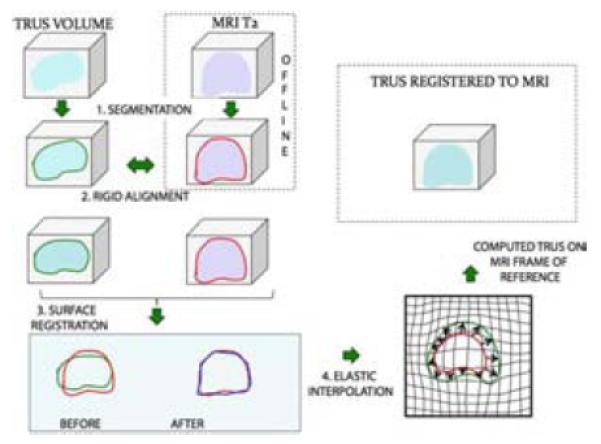
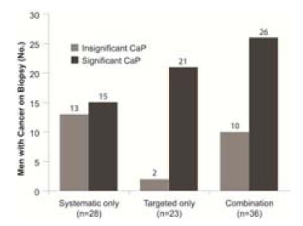
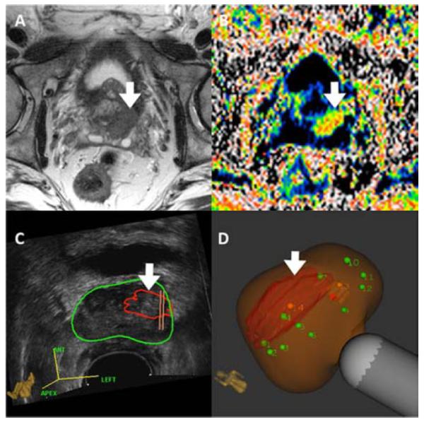
Multiparametric magnetic resonance imaging (MRI) has provided a method for visualizing prostate cancer. MRI-ultrasonography fusion allows prostate biopsy to be performed quickly, on an outpatient basis, using the transrectal technique. Targeted biopsies are more sensitive for detection of prostate cancer than nontargeted, systematic biopsies and detect more significant prostate cancers and fewer insignificant cancers than conventional biopsies. A negative MRI scan should not defer biopsy. Two groups who will especially benefit from targeted prostate biopsy are men with low-risk lesions in active surveillance and men with increased prostate-specific antigen levels and previous negative conventional biopsies.

Keywords: Fusion biopsy; Magnetic resonance imaging; Prostate cancer; Prostate-specific antigen; Targeted prostate biopsy; Transrectal ultrasonography; Ultrasonography.


Figures

Figure 1
Figure 1
Prostate MRI c. 1983. Among the first published MRI images were those above, obtained with a 0.35T coil. In the transverse scan (A), the prostate (P) is enlarged and the Foley catheter (arrow) in the prostatic urethra is displaced posteriorly to the left by adenomatous tissue. Seminal vesicles are seen inferior to the bladder (s). In the sagittal scan (B), air (A) and urine (U) level can be seen in the bladder. At the time, magnet strength was not capable of demonstrating zonal anatomy or small cancers.From Hricak H, Williams RD, Spring DB, et al. Anatomy and pathology of the male pelvis by magnetic resonance imaging. AJR. American journal of roentgenology. Dec 1983;141(6):1101-1110; with permission.
Figure 2
Figure 2
CaP visualized by multi-parametric MRI (mp-MRI). Arrows point to lesion.(A) T2-weighted image, (B)diffusion weighted imaging (DWI), (C)dynamic contrast enhancement (DCE), (D) whole mount specimen obtained by radical prostatectomy, showing cancer.From Natarajan S, Marks LS, Margolis DJ, et al. Clinical application of a 3D ultrasound-guided prostate biopsy system. Urologic oncology. May-Jun 2011;29(3):334-342; with permission.
Figure 3
Figure 3
Process of MRI-US Fusion. MR and TRUS images are outlined or segmented (1) and then rigidly aligned (2). Fusion then proceeds involving a surface registration (3), and elastic (non-rigid) interpolation (4). Finally, the registered, or superimposed images are produced on a monitor, where targeted biopsy is performed. The target is derived from the MRI; the biopsy aiming is via real-time ultrasound.From Natarajan S, Marks LS, Margolis DJ, et al. Clinical application of a 3D ultrasound-guided prostate biopsy system. Urologic oncology. May-Jun 2011;29(3):334-342; with permission.
Figure 4
Figure 4
In-Bore MRI guided biopsy is performed prone; patient undergoes a diagnostic MRI in advance of biopsy; the images are then processed and delineated; patient subsequently returns to MR facility for procedure, which involves fusing the diagnostic MRI with the 2nd MRI used to guide biopsy. Courtesy of Invivo Corporation, Gainesville, FL; with permission.
Figure 5
Figure 5. UroNav Fusion Device
Originally developed in a collaboration between Phillips and the National Cancer Institute, the UroNav system uses an external magnetic field generator (A) for tracking a biopsy needle’s position in 3D space which is recorded at an imaging terminal (B). Courtesy of Invivo Corporation, Gainesville, FL; with permission.
Figure 6
Figure 6. Artemis Fusion Device
Originally developed at Robarts Research Institute in Canada, the Artemis device gained FDA-approved in 2008. It is manufactured by Eigen (Grass Valley, Ca). The Artemis device uses a mechanical arm with built-in encoders to track biopsy location. During atransrectal ultrasound scan, 2D images are digitized with a frame grabber and reconstructed into a 3D image.A model of the prostate is then generated from the 3D image; biopsy, tracking of the biopsy site, and MRI fusion are then performed on the reconstructed model. Courtesy of Eigen, Grass Valley, CA; with permission.
Figure 7
Figure 7. Tracking Arm Encoders
(A) Prototype created at Robarts Research Institute, (B) Working model in current use. Arrows denote location of the 3 encoders. As the TRUS transducer and cradle are moved, encoders (arrows) in the tracking mechanism measure the angles between linkages, and software calculates the transducer tip position and orientation in real-time.From Bax J, Cool D, Gardi L, et al. Mechanically assisted 3D ultrasound guided prostate biopsy system. Med Phys. Dec 2008;35(12):5397-5410; with permission.
Figure 8
Figure 8. Biopsy Results for Patients with Prior Negative Systematic Biopsies
This chart shows the number of subjects diagnosed with significant cancers (dark grey) and insignificant cancers (light grey), depending on biopsy method. Clinically significant cancer was defined as Gleason >6 or >4 mm maximal core length .Targeted biopsy detected more significant cancers and fewer insignificant cancers than systematic biopsy. 15 patients were diagnosed only by systematic biopsy, i.e., cancer was present in areas where the MRI showed no abnormality. The false negative rate of MRI is not yet known. From Sonn GA, Chang E, Natarajan S, et al. Value of Targeted Prostate Biopsy Using Magnetic Resonance-Ultrasound Fusion in Men with Prior Negative Biopsy and Elevated Prostate-specific Antigen. Eur Urol. Mar 17 2013; with permission.
Figure 9
Figure 9
Patient with two prior negative biopsies wasfound to have a suspicious regionon MRI (Image Score =5) and underwent targeted confirmatory biopsy via MRI-US fusion. Gleason 3+4=7 CaP was found in the anterior target. A – T2-weighted MR image, B – Colorized apparent diffusion coefficient (ADC) MR image, C- Ultrasound showing prostate contour with areas of suspicion outlined (large grade 5 target, arrow), D – 3D reconstructed model of prostate showing targets and biopsy cores (tan lines).
Figure 10
Figure 10
mpMRI(Panels A& B) and Artemis images (Panels C & D) of prostate from 64y.o. Caucasian male, who was enrolled in active surveillance on the basis of a microfocal lesion on conventional biopsy. Panel A shows T2WI. Panel B shows DWI. Panel C shows the lesion outlined in red, superimposed on the ultrasound image of the prostate (green circle).Panel D shows the lesion after MRI-US fusion; green dots are sites for systematic biopsy.Cancer was found only on targeted, but not systematic biopsies. Defining tumor burden in men with apparent ‘low risk’ CaP is an important use of targeted biopsy.

References

    1. Silletti JP, Gordon GJ, Bueno R, Jaklitsch M, Loughlin KR. Prostate biopsy: past, present, and future. Urology. 2007 Mar;69(3):413–416. - PubMed
    1. Hodge KK, McNeal JE, Terris MK, Stamey TA. Random systematic versus directed ultrasound guided transrectal core biopsies of the prostate. The Journal of urology. 1989 Jul;142(1):71–74. discussion 74-75. - PubMed
    1. Cooperberg MR, Broering JM, Kantoff PW, Carroll PR. Contemporary trends in low risk prostate cancer: risk assessment and treatment. J Urol. 2007 Sep;178(3):S14–19. Pt 2. - PMC - PubMed
    1. King CR, McNeal JE, Gill H, Presti JC., Jr. Extended prostate biopsy scheme improves reliability of Gleason grading: implications for radiotherapy patients. Int J Radiat Oncol Biol Phys. 2004 Jun 1;59(2):386–391. - PubMed
    1. Hricak H, Williams RD, Spring DB, et al. Anatomy and pathology of the male pelvis by magnetic resonance imaging. AJR. American journal of roentgenology. 1983 Dec;141(6):1101–1110. - PubMed

Publication types