. 2014 Apr 12:14:48.
doi: 10.1186/1471-2261-14-48.

The prevalence of coronary anomalies in a single center of Korea: origination, course, and termination anomalies of aberrant coronary arteries detected by ECG-gated cardiac MDCT

Affiliations

The prevalence of coronary anomalies in a single center of Korea: origination, course, and termination anomalies of aberrant coronary arteries detected by ECG-gated cardiac MDCT

June Namgung et al. BMC Cardiovasc Disord. .

Abstract

Background: Coronary anomalies are rare congenital abnormalities often found incidentally on conventional coronary angiography (CCA) or coronary CT angiography (CTA). They may result in various clinical outcomes. CCA is invasive and not able to demonstrate all coronary anomalies in detail, especially those with complex courses. Multidetector computed tomography (MDCT) enables visualization of the origin and course of coronary arteries. The objective of this study was to investigate the prevalence of origin and termination coronary artery anomalies and the course of these anomalies in patients in a single center in Korea.

Methods: To diagnose coronary anomalies, the angiographic data of 8,864 consecutive patients undergoing 64- or 320-MDCT from September 2005 to November 2011 were analyzed retrospectively.

Results: Among the 8,864 patients, 103 (1.16%) had coronary anomalies. Ninety (87.4%) patients had origin and distribution anomalies, and 13 (12.6%) patients had a coronary artery fistula. The most common anomaly (41, 39.8%) was an anomalous origin of the right coronary artery (RCA). Of these, three patients received a coronary artery bypass graft.

Conclusions: The prevalence of coronary anomalies in a single center of Korea was 1.16%. The incidence and patterns of coronary artery anomalies in our patient population were similar to those of previous studies.


Figures

Figure 1
Figure 1
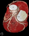
Anomalous origin of the right coronary artery (RCA) originating from the left coronary sinus of Valsalva (LSV). 3D volume-rendered coronary image (A) and curved, multiplanar reconstruction image (B) showing the anomalous origin of the right coronary artery (black arrow) from the LCS, and courses between the aorta (Ao) and the pulmonary artery (PA). Cross-sectional image (C) showing the proximal RCA, with narrowing and lateral compression resulting in an ovoid lumen.
Figure 2
Figure 2
Origin and course of the anomalies of the left main coronary artery (LMCA) from the right sinus of Valsalva (RSV). Volume rendering image shows the LMCA arising from the RSV (A). It depicts the long LMCA branching into the left circumflex artery at the proximal interventricular groove and the RCA arising normally from the RSV. The curved multiplanar reconstruction image (B) shows the opening of the LMCA, acute angle take-off of the RSV from the aorta, and the interarterial course between the ascending aorta and the pulmonary artery (PA).
Figure 3
Figure 3
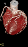
Anomalous origin of the left circumflex artery (LCX) from the right sinus of Valsalva (RSV). Volume rendering image shows the LCX arising from the RSV (A) or the right coronary artery (B), and travelling between the aorta and the left atrium (retroaortic course).
Figure 4
Figure 4
Absent left main trunk (split origin of left coronary artery). 3D volume rendering image shows absent left main coronary artery (LMCA) and the separate origin of the left anterior descending coronary artery (LAD) and left circumflex artery (LCX).
Figure 5
Figure 5
Volume rendering image of single coronary ostium in the left sinus of Valsalva (LSV). The dilated left main coronary artery (LMCA) divided into the left anterior descending coronary artery (LAD) and the left circumflex artery (LCX). The LCS then coursed in the left atrioventricular groove and continued to the posterior atrioventricular groove where it occupied the anatomic position normally occupied by the right coronary artery (RCA).
Figure 6
Figure 6
High takeoff of the right coronary artery (RCA). The volume rendering image shows the origin of the RCA from the anterior surface of the ascending aorta approximately 2 cm above the sinotubular junction. The first portion of the proximal RCA shows a hockey-stick type of configuration, with an acute-angle bend on its main stem, before returning to its regular course in the right atrioventricular groove.
Figure 7
Figure 7
Duplication of the left anterior descending coronary artery (LAD). A: 3D reconstruction, showing the course of the long LAD from its origin in the right coronary artery (RCA), then passing anterior to the pulmonary artery (PA) and following a course to the anterior interventricular groove where it follows the course of a traditional distal LAD. The short LAD courses and terminates in the anterior interventricular sulcus, without reaching the apex. B: 3D reconstruction showing the left main coronary artery (LMCA) and its short LAD branching the diagonal branches.

References

    1. Yamanaka O, Hobbs RE. Coronary artery anomalies in 126,595 patients undergoing coronary angiography. Cathet Cardiovasc Diagn. 1990;21(1):28–40. doi: 10.1002/ccd.1810210110. - DOI - PubMed
    1. Kaku B, Shimizu M, Yoshio H, Ino Hidkazu, Mizuno S, Kanaya H, Ishise S, Mabuchi H. Clinical features and prognosis of Japanese patients with anomalous origin of the coronary artery. Jpn Circ J. 1996;60(10):731–741. doi: 10.1253/jcj.60.731. - DOI - PubMed
    1. Kardos A, Babai L, Rudas L, Gaál T, Horváth T, Tálosi L, Tóth K, Sárváry L, Szász K. Epidemiology of congenital coronary artery anomalies: a coronary arteriography study on a central European population. Cathet Cardiovasc Diagn. 1997;42(3):270–275. doi: 10.1002/(SICI)1097-0304(199711)42:3<270::AID-CCD8>3.0.CO;2-9. - DOI - PubMed
    1. Garg N, Tewari S, Kapoor A, Gupta DK, Sinha N. Primary congenital anomalies of the coronary arteries: a coronary arteriographic study. Int J Cardiol. 2000;74(1):39–46. doi: 10.1016/S0167-5273(00)00243-6. - DOI - PubMed
    1. Yildiz A, Okcun B, Peker T, Arslan C, Olcay A, Bulent Vatan M. Prevalence of coronary artery anomalies in 12,457 adult patients who underwent coronary angiography. Clin Cardiol. 2010;33(12):E60–E64. doi: 10.1002/clc.20588. - DOI - PMC - PubMed

MeSH terms

LinkOut - more resources