

Unilateral morning glory optic disc anomaly in a case with Down syndrome
- PMID: 24725623
- PMCID: PMC3989808
- DOI: 10.1186/1471-2415-14-48
Unilateral morning glory optic disc anomaly in a case with Down syndrome
Abstract
Background: This case is unique because it is the first reported case of Down syndrome with morning glory optic disc anomaly in literature.
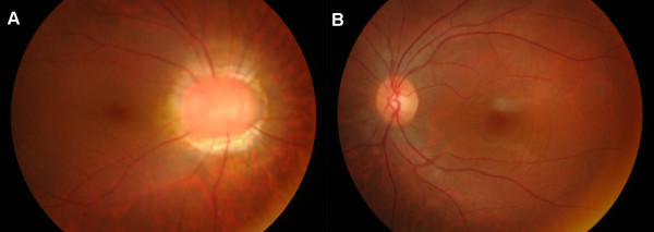
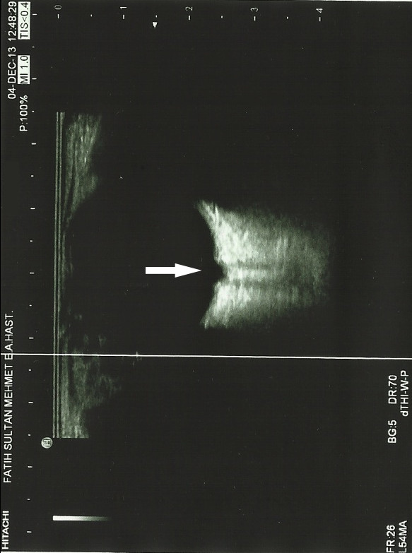
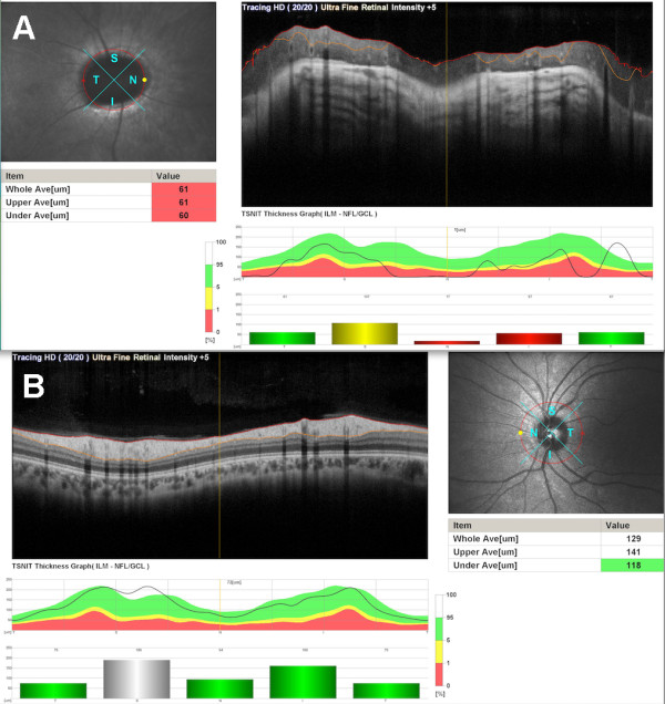
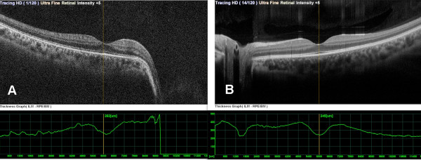
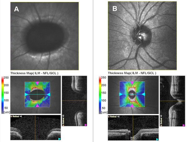
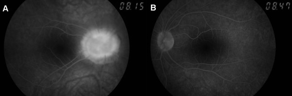
Case presentation: A 15-year-old girl with features of Down syndrome presented to the Clinic of Ophthalmology for a regular ophthalmologic examination. Her best corrected visual acuity was 20/50 in the right eye and 20/20 in the left eye. The fundus examination revealed findings compatible with unilateral morning glory optic disc anomaly in the right eye. The patient underwent a complete ophthalmologic and systemic evaluation to explore possible associated findings.
Conclusion: This case report emphasizes the importance of ophthalmic screening-examinations in Down children to rule out any vision relevant pathology.
Figures

References
-
- Down JL. Observations on an ethnic classification of idiots. Ment Retard. 1995;33(1):54–56. - PubMed
-
- Improved national prevalence estimates for 18 selected major birth defects--United States, 1999-2001. MMWR Morb Mortal Wkly Rep. 2006;54(51):1301–1305. - PubMed
-
- Davis JS. Ocular manifestations in Down syndrome. Pa Med. 1996;99(suppl):67–70. - PubMed
-
- Kindler P. Morning glory syndrome: unusual congenital optic disk anomaly. Am J Ophthalmol. 1970;69:376–384. - PubMed
Publication types
MeSH terms
LinkOut - more resources
Full Text Sources
Other Literature Sources
Medical
