. 2014 Apr:454-455:78-92.
doi: 10.1016/j.virol.2014.01.030. Epub 2014 Feb 25.

Lack of group X secreted phospholipase A₂ increases survival following pandemic H1N1 influenza infection

Affiliations

Lack of group X secreted phospholipase A₂ increases survival following pandemic H1N1 influenza infection

Alyson A Kelvin et al. Virology. 2014 Apr.

Abstract

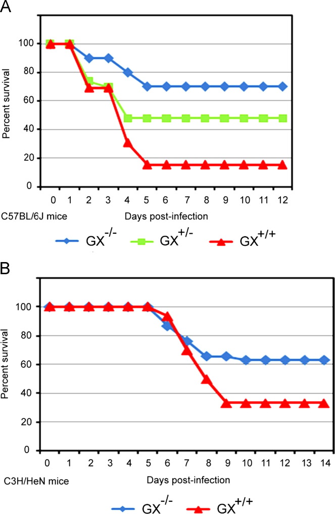
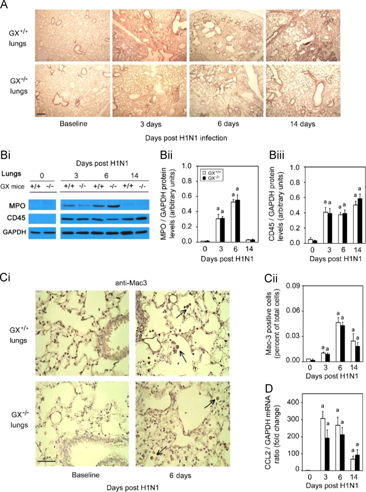
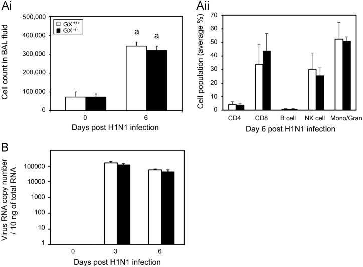
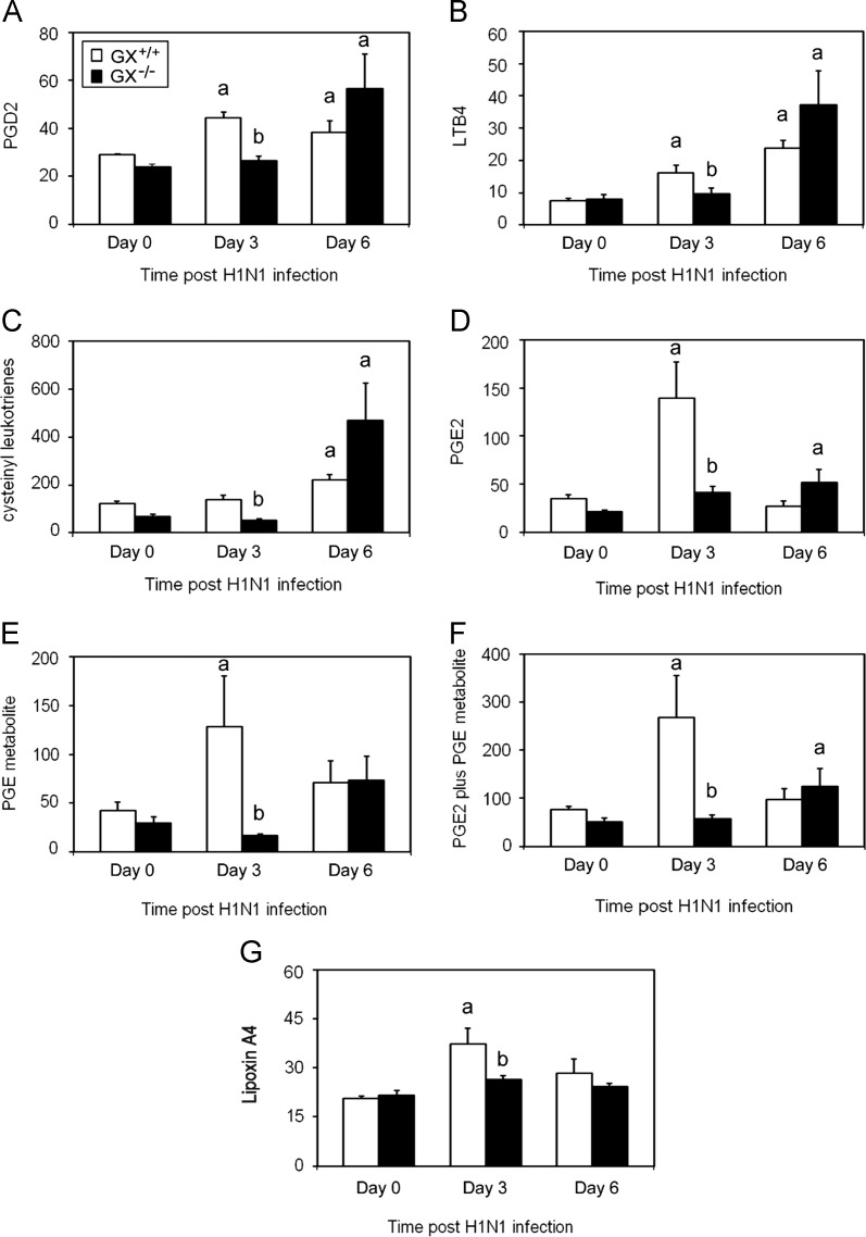
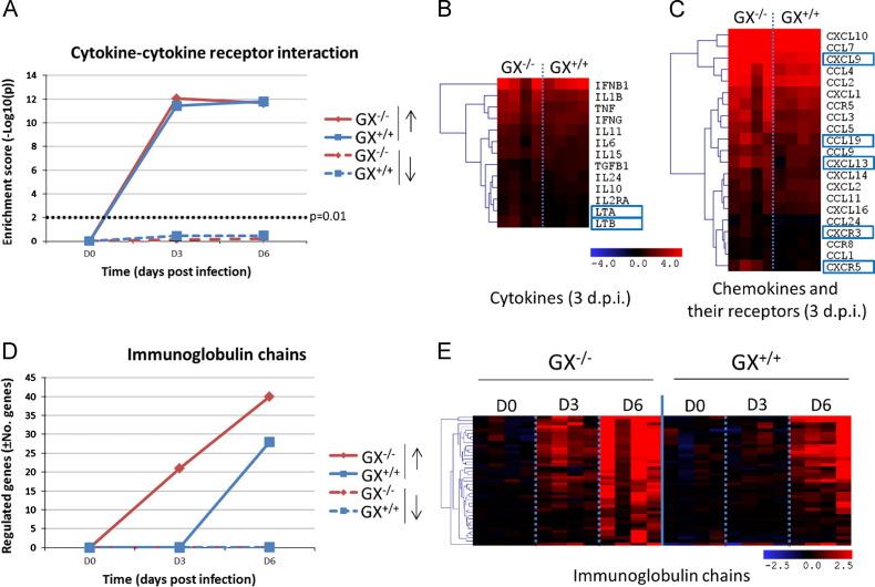
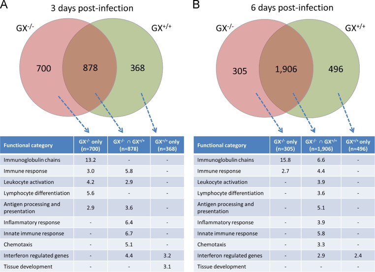
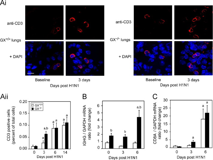
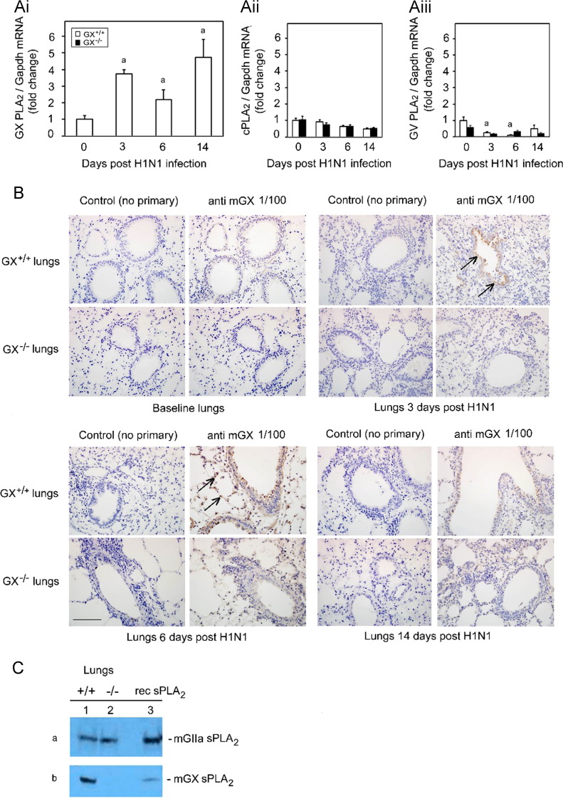
The role of Group X secreted phospholipase A2 (GX-sPLA2) during influenza infection has not been previously investigated. We examined the role of GX-sPLA2 during H1N1 pandemic influenza infection in a GX-sPLA2 gene targeted mouse (GX(-/-)) model and found that survival after infection was significantly greater in GX(-/-) mice than in GX(+/+) mice. Downstream products of GX-sPLA2 activity, PGD2, PGE2, LTB4, cysteinyl leukotrienes and Lipoxin A4 were significantly lower in GX(-/-) mice BAL fluid. Lung microarray analysis identified an earlier and more robust induction of T and B cell associated genes in GX(-/-) mice. Based on the central role of sPLA2 enzymes as key initiators of inflammatory processes, we propose that activation of GX-sPLA2 during H1N1pdm infection is an early step of pulmonary inflammation and its inhibition increases adaptive immunity and improves survival. Our findings suggest that GX-sPLA2 may be a potential therapeutic target during influenza.

Keywords: H1N1 pandemic influenza; Host response; Inflammation; Influenza; Leukotrienes; Lipoxin A(4); Pathogenesis; Phospholipids; Prostaglandins; Secreted phospholipase A(2).


Figures

Fig. 2
Fig. 2
Increased survival of GX−/− vs. GX+/− or GX+/+ mice following A/Mexico/4108/2009 infection. GX+/+ (n=25), GX+/− (n=32), and GX−/− (n=24) mice (C57BL/6J background, lacks GIIA-sPLA2) were infected intranasally with A/Mexico/4108/2009 and survival was assessed for a 14 day period (A). GX+/+ (n=71) and GX−/− (n=57) mice (C3H/HeN background, expresses GIIA-sPLA2) were infected intranasally with A/Mexico/4108/2009 and survival was assessed for a 14 day period (B). Animals were sacrificed if their body weight decreased to less than 80% of baseline weight, or if the 14-day duration of the study was completed. Log rank test, p<0.05 GX−/− vs. GX+/+ and GX+/− mice or p<0.05, GX−/− vs. GX+/+ mice. All the mice used in these experiments were genotyped littermates and grouped and analyzed by their genotype.
Fig. 3
Fig. 3
Infection with H1N1pdm influenza induces similar pulmonary inflammation and recruitment of inflammatory cells in GX+/+ and GX−/−mice. GX+/+ and GX−/− mice (C3H/HeN background mice) were infected with H1N1pdm (A/Mexico/4108/2009) influenza and the lungs were perfusion fixed in situ with 4% paraformaldehyde on specific time points following infection, sectioned and subject to hematoxylin and eosin staining (A). MPO protein (neutrophil marker), CD45 protein (leukocyte marker) and GAPDH protein (loading control) expression levels were determined by immunoblot analysis from lung tissue homogenates of GX+/+ and GX−/− mice over a 14 day time course of H1N1pdm influenza infection (Bi). Densitometric analysis of MPO (Bii) and CD45 (Biii) protein levels normalized to GAPDH levels in lung tissue of GX+/+ (open bars) and GX−/− (filled bars) mice after H1N1pdm influenza infection, are presented. Immunohistochemical analysis with specific rabbit primary antibody against mouse Mac-3 antigen (marker for macrophages) is shown (Ci). Assessment of Mac-3 positive cells (indicated by →) per high power field before, 3, 6 or 14 days after infection with H1N1pdm influenza is shown (Cii). CCL2 mRNA expression normalized to GAPDH was determined by quantitative real-time PCR in lung tissue of GX+/+ and GX−/− mice after H1N1pdm influenza infection (D). Representative images (×200) from five independent experiments are shown. Scale bar: 100 μm (A) or 50 μm (C). a, p<0.05 GX+/+ or GX−/− vs. base; b, p<0.05 GX+/+ vs. GX−/− at any time point, ANOVA followed by paired t-test, two tailed, assuming unequal variance. n≥8 per group.
Fig. 4
Fig. 4
Lung viral titers and cell counts in BAL fluid show similar cell numbers and cell population distributions following infection in GX+/+ and GX−/− mice. GX−/− and GX+/+ mice infected with A/Mexico/4108/2009 were investigated for lung cell numbers, populations and viral load. BAL fluid was harvested from infected GX+/+ (open bars) and GX−/− (filled bars) mice (C3H/HeN background mice) on day 0 and 6 and the cell numbers (Ai) and cell population distributions (Aii) were assessed by FACS. Viral load was determined on day 0, 3 and 6 pi of GX+/+ (open bars) and GX−/− (filled bars) mice by Real-Time RT-PCR vRNA quantification (B). All the mice used in these experiments were genotyped littermates and grouped and analyzed by their genotype. a, p<0.05 GX+/+ vs. base, ANOVA followed by paired t-test, two tailed, assuming unequal variance. n≥7 per group.
Fig. 5
Fig. 5
Decreased eicosanoid levels in the BAL fluid 3 but not 6 days after infection with A/Mexico/4108/2009 in GX−/− vs. GX+/+ mice. The eicosanoid levels in the BAL fluid of GX−/− and GX+/+ mice (C3H/HeN background) were investigated at baseline and following infection with A/Mexico/4108/2009. GX+/+ (open bars) and GX−/− (filled bars) mice (C3H/HeN background mice) BAL fluid was harvested by instilling ice cold NaCl (1 ml) five times and pooled. Levels of PGD2 MOX (A), LTB4 (B), cysteinyl leukotrienes (C), PGE2 (D), stable PGE metabolite (E), PGE2 plus PGE metabolite (F) and Lipoxin A4 (G) were assessed by ELISA. These results are the mean of 5 independent studies. All the mice used in these experiments were genotyped littermates and grouped and analyzed by their genotype. GX+/+ (open bars) and GX−/− (filled bars). Results are expressed in pg/mL. a, p<0.05 GX+/+ or GX−/− vs. base; b, p<0.05 GX+/+ vs. GX−/− at any time point, ANOVA followed by paired t-test, two tailed, assuming unequal variance. n≥7 per group.
Fig. 6
Fig. 6
Effect of GX-sPLA2 deficiency in the mRNA expression levels of cytokines, chemokines and their receptors and immunoglobulin chains in the lung tissue of mice during influenza infection. GX+/+ and GX−/− mice (C3H/HeN background mice) were infected with influenza A/Mexico/4108/2009 and the gene expression profiles were analyzed in the lung tissue at day 0, 3, and 6 days after infection by microarray analysis (n=4 per group). Evolution of gene enrichment (Fisher׳s exact test) of the KEGG category “cytokine-cytokine receptor interaction” (A). Differences in the expression levels of cytokines (B) and chemokines (C) and their receptors at 3 days pi. The heatmaps show the genes that are significantly upregulated with respect to the control group and the blue boxes indicate that the expression levels of those genes are significantly higher in the GX−/− than in the wild type mice at the same time-point. Evolution in the expression levels of immunoglobulin genes: total number of regulated genes (D) and overview of different experimental groups and time-points (E). All the mice used in these experiments were genotyped littermates and grouped and analyzed by their genotype.
Fig. 7
Fig. 7
Intersect analysis of the genes up-regulated in the lung tissue of GX−/− and GX+/+ mice during influenza infection and functional classification of the resulting gene subsets. Venn diagrams are representative of the total number of genes that are significantly up-regulated with respected to the uninfected mice. David Annotation tool was used to classify the genes of each subset, and the fold enrichment is shown for each category. All the mice used in these experiments were genotyped littermates and grouped and analyzed by their genotype. * The “Immunoglobulin chains” category was manually curated and contains 84 genes. ⁎⁎ The “interferon responses category”.
Fig. 8
Fig. 8
GX-sPLA2 deficiency increases T cell recruitment and immunoglobulin heavy chain mRNA expression in the lung tissue of mice during influenza infection. Day 0 and 3 pi with H1N1pdm (A/Mexico/4108/2009) influenza the lungs from GX+/+ and GX−/− mice (C3H/HeN background mice) were perfusion fixed in situ with 4% paraformaldehyde, sectioned and subject to immunofluorescence analysis with specific rabbit primary antibody against mouse CD3 antigen (marker for T-cell) (Ai). Representative images (at ×400 with 3.4 zoom factor) from five independent experiments are shown. Assessment of CD3 positive cells per high power field for day 0, 3, 6 and 14 days following infection with H1N1pdm influenza is shown (Aii). IgG (B) and CD8A (C) mRNA expression normalized to GAPDH were determined by quantitative real-time PCR in lung tissue of GX+/+ and GX−/− mice after H1N1pdm influenza infection. Scale bar: 10 μm. a, p<0.05 GX+/+ or GX−/− vs. base; b, p<0.05 GX+/+ vs. GX−/− at any time point, ANOVA followed by paired t-test, two tailed, assuming unequal variance. n≥8 per group. All the mice used in these experiments were genotyped littermates and grouped and analyzed by their genotype.
Fig. 1
Fig. 1
Infection with H1N1pdm influenza stimulates the expression of GX-sPLA2 in bronchial epithelial cells and inflammatory cells. Mice were infected with H1N1pdm (A/Mexico/4108/2009) and the lungs were assessed for the mRNA and protein expression and localization of PLAs during a 14 day time course. GX-sPLA2 mRNA (Ai), cPLA2 mRNA (Aii) and GV-sPLA2 mRNA (Aiii) expression quantified by Real-Time RT-PCR was normalized to GAPDH, GX+/+ (open bars) and GX−/− (filled bars) mice (C3H/HeN background mice). GX+/+ and GX−/− mouse lungs were perfusion fixed in situ with 4% paraformaldehyde, sectioned and subject to immunohistochemical analysis with the IgG fraction of rabbit anti-mouse GX-sPLA2 antiserum (1/100 dilution) (B). GIIA and GX-sPLA2 protein expression determined by immunoblot analysis of lung tissue homogenates of wild type GX+/+ (lane 1) and knockout GX−/− (lane 2) mice (C). For each blot, the corresponding recombinant sPLA2 enzyme (rec sPLA2) was run alone (lane 3) as a control. Representative results for five separate experiments are shown. All the mice used in these experiments were genotyped littermates and grouped and analyzed by their genotype. a, p<0.05 GX+/+ or GX−/− vs. base; b, p<0.05 GX+/+ vs. GX−/− at any time point, ANOVA followed by paired t-test, two tailed, assuming unequal variance. n≥8 per group; 400×; scale bar: 50 μm for immunohistochemistry.

References

    1. Arima M., Fukuda T. Prostaglandin D(2) and T(H)2 inflammation in the pathogenesis of bronchial asthma. Korean J. Intern. Med. 2011;28:8–18. - PMC - PubMed
    1. Baillie J.K., Digard P. Influenza – time to target the host? N. Engl. J. Med. 2013;369:191–193. - PubMed
    1. Bermejo-Martin J.F., Ortiz de L.R., Pumarola T., Rello J., Almansa R., Ramirez P., Martin-Loeches I., Varillas D., Gallegos M.C., Seron C., Micheloud D., Gomez J.M., Tenorio-Abreu A., Ramos M.J., Molina M.L., Huidobro S., Sanchez E., Gordon M., Fernandez V., Del C.A., Marcos M.A., Villanueva B., Lopez C.J., Rodriguez-Dominguez M., Galan J.C., Canton R., Lietor A., Rojo S., Eiros J.M., Hinojosa C., Gonzalez I., Torner N., Banner D., Leon A., Cuesta P., Rowe T., Kelvin D.J. Th1 and Th17 hypercytokinemia as early host response signature in severe pandemic influenza. Crit. Care. 2009;13:R201. (doi:cc8208 [pii];10.1186/cc8208 [doi]) - PMC - PubMed
    1. Bermejo-Martin J.F., Martin-Loeches I., Rello J., Anton A., Almansa R., Xu L., Lopez-Campos G., Pumarola T., Ran L., Ramirez P., Banner D., Ng D.C., Socias L., Loza A., Andaluz D., Maravi E., Gomez-Sanchez M.J., Gordon M., Gallegos M.C., Fernandez V., Aldunate S., Leon C., Merino P., Blanco J., Martin-Sanchez F., Rico L., Varillas D., Iglesias V., Marcos M.A., Gandia F., Bobillo F., Nogueira B., Rojo S., Resino S., Castro C., Ortiz de L.R., Kelvin D. Host adaptive immunity deficiency in severe pandemic influenza. Crit. Care. 2010;14:R167. (doi:cc9259 [pii];10.1186/cc9259 [doi]) - PMC - PubMed
    1. Bhavsar P.K., Levy B.D., Hew M.J., Pfeffer M.A., Kazani S., Israel E., Chung K.F. Corticosteroid suppression of lipoxin A4 and leukotriene B4 from alveolar macrophages in severe asthma 12. Respir. Res. 2010;11:71. (doi:1465-9921-11-71 [pii];10.1186/1465-9921-11-71 [doi]) - PMC - PubMed

Publication types

Substances

LinkOut - more resources