. 2014 Apr:454-455:157-68.
doi: 10.1016/j.virol.2014.02.005. Epub 2014 Mar 4.

Phagocytic cells contribute to the antibody-mediated elimination of pulmonary-infected SARS coronavirus

Affiliations

Phagocytic cells contribute to the antibody-mediated elimination of pulmonary-infected SARS coronavirus

Fumihiko Yasui et al. Virology. 2014 Apr.

Erratum in

Abstract

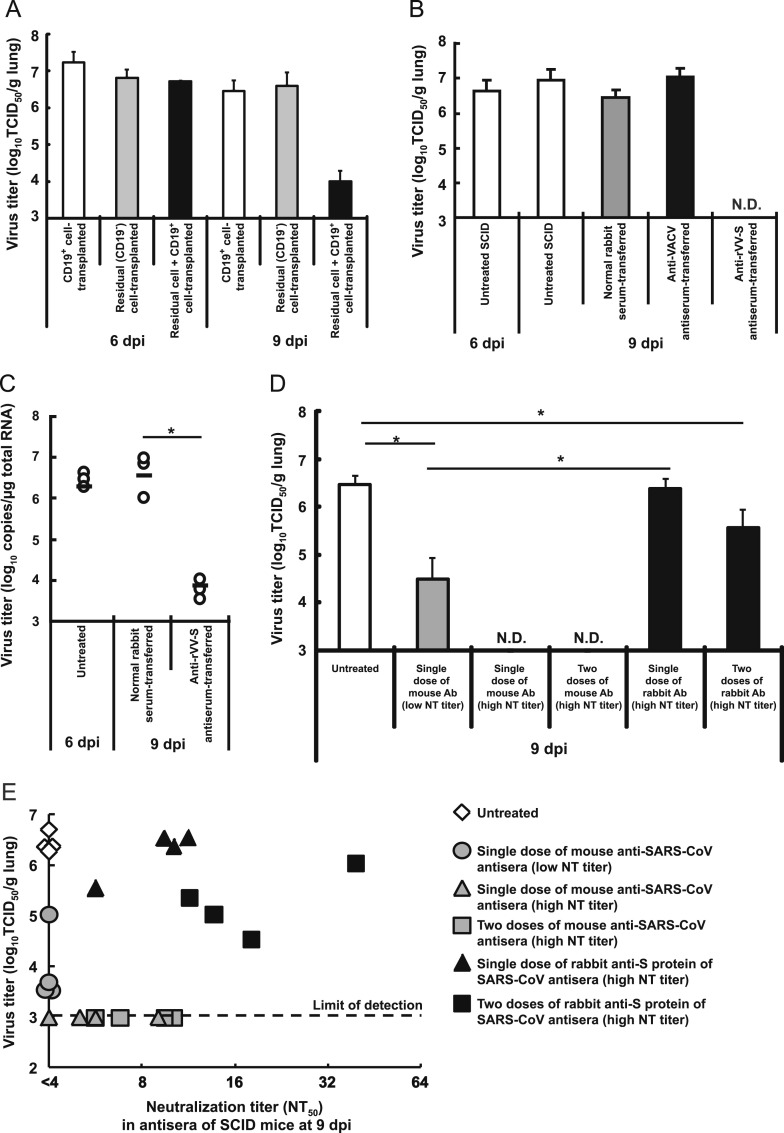
While the 2002-2003 outbreak of severe acute respiratory syndrome (SARS) resulted in 774 deaths, patients who were affected with mild pulmonary symptoms successfully recovered. The objective of the present work was to identify, using SARS coronavirus (SARS-CoV) mouse infection models, immune factors responsible for clearing of the virus. The elimination of pulmonary SARS-CoV infection required the activation of B cells by CD4(+) T cells. Furthermore, passive immunization (post-infection) with homologous (murine) anti-SARS-CoV antiserum showed greater elimination efficacy against SARS-CoV than that with heterologous (rabbit) antiserum, despite the use of equivalent titers of neutralizing antibodies. This distinction was mediated by mouse phagocytic cells (monocyte-derived infiltrating macrophages and partially alveolar macrophages, but not neutrophils), as demonstrated both by adoptive transfer from donors and by immunological depletion of selected cell types. These results indicate that the cooperation of anti-SARS-CoV antibodies and phagocytic cells plays an important role in the elimination of SARS-CoV.

Keywords: Antibody; B cells; CD4(+) T cells; Elimination; Phagocytic cells; SARS-CoV.


Figures

Fig. 1
Fig. 1
CD4+ T cells play an important role in the control of SARS-CoV infection. (A–C) aged BALB/c mice (n=4–7/time point), young BALB/c mice (n=5–10/time point), and young SCID mice (n=4–8/time point) were infected intranasally with 1×10⁵ TCID50 of SARS-CoV Vietnam strain. (A) Virus titers in the lung (TCID50/g lung tissue) of aged BALB/c mice (closed circles), young BALB/c mice (gray circles), or young SCID mice (open squares) sacrificed at 2, 4, 6, 9, or 21 dpi (except for 20 dpi in young BALB/c mice). p<0.05, ⁎⁎p<0.01 (compared with young BALB/c mice and SCID mice at the respective time point). N.D.: not detected. (B) Representative lung sections (hematoxylin and eosin staining; section thickness 4 μm) from aged BALB/c mice at 9 dpi (a and c) and 21 dpi (b and d) and from young SCID mice at 9 dpi (e and g) and 21 dpi (f and h). For all micrographs, original magnification is 200×. (C) Detection of virus-infected cells in the lungs at 2, 9, or 21 dpi (SARS-CoV nucleocapsid protein [brown staining]; original magnification, 400×). (D) Temporal change of pulmonary virus titer in the following: aged BALB/c mice (green); untreated SCID mice (blue); SCID mice transplanted with splenocytes from naïve BALB/c mice (yellow); or SCID mice transplanted with splenocytes from sensitized BALB/c mice (red). Splenocytes (4×10⁷ cells) were administered intravenously to each recipient SCID mouse 1 day before infection. Data are presented as mean±S.D. (n=4/time point). p<0.05 (compared with naïve splenocyte-transplanted SCID mice at 2 dpi or with other groups at 4 dpi). (E) Representative lung sections (hematoxylin and eosin staining; section thickness 4 μm) from each group in (D) at 9 dpi. SPL, splenocyte. For all micrographs, original magnification is 200×. (F) Flow cytometry analysis of CD4 and CD8 expression on lymphocytes isolated from spleen 1 day after administration of the indicated mAb. (G) Virus titers in the lung of untreated (white), CD8+ cell-depleted (light gray), CD4+ cell-depleted (dark gray), or CD4+ and CD8+ cell-depleted BALB/c mice (black) at 6 and 9 dpi. The limit of detection was <1×10³ TCID50/g lung. Data are presented as mean±S.D. (n=3–7/time point).
Fig. 2
Fig. 2
Antiviral effect of CD4+ cells is indirect. (A) Temporal change of virus titer in the lung of young BALB/c mice (closed circles) and young nude mice (open circles). Data are presented as mean±S.D. (n=5/time point). N.D.: not detected. (B) Representative lung sections (hematoxylin and eosin staining; section thickness 4 μm) from young BALB/c mice at 9 dpi (a) and 20 dpi (b) and young nude mice at 9 dpi (c) and 20 dpi (d). For all micrographs, original magnification is 200×. (C) Virus titer in the lung of CD4+ cell-transplanted SCID mice was determined at 6 and 9 dpi. Data are presented as mean±S.D. (n=5/time point). CD4+ cells (1×10⁷ cells) were administered to each recipient SCID mouse intravenously 1 day before challenge. (D) Virus titer in the lung of nude mice adoptively transplanted with CD4+ cells (gray) or residual cells (CD4 cells; black), determined at 6 and 9 dpi. Untreated nude mice (white) were included as a control. CD4+ cells (1×10⁷ cells) or residual cells (3×10⁷ cells) were administered to each recipient nude mouse intravenously 1 day before challenge. Data are presented as mean±S.D. (n=3/time point). (E) Pulmonary virus titer of IFN-γ deficient mice and splenocyte-transplanted SCID mice at 6 and 9 dpi. Splenocytes (4×10⁷ cells) derived from IFN-γ deficient mice were administered to each recipient SCID mouse intravenously 1 day before challenge. Data are presented as mean±S.D. (n=3/time point). N.D.: not detected. SPL, splenocyte.
Fig. 3
Fig. 3
Clearance of SARS-CoV with anti-SARS-CoV antibodies does not correlate with neutralizing activity. (A) Virus titers in the lung of SCID mice adoptively transplanted with CD19+ cells (white), residual cells (CD19 cells; gray), or both CD19+ and CD19 cells (black) were determined at 6 and 9 dpi. CD19+ cells (2×10⁷ cells) and/or residual cells (2×10⁷ cells) were administered to each recipient SCID mouse intravenously 1 day before challenge. Data are presented as mean±S.D. (n=3–4 mice/time point). (B) Antiviral effect of post-infection administration of rabbit anti-S (spike) protein of SARS-CoV antibody (NT50>300) (at 6 and 8 dpi) assessed as pulmonary virus titer at 9 dpi; untreated (white), normal rabbit serum-injected (gray), anti-vaccinia virus (VACV) antiserum (black), or anti-recombinant vaccinia virus expressing S protein of SARS-CoV (rVV-S) antiserum-injected (anti-S protein of SARS-CoV antiserum-injected). Data are presented as mean±S.D. (n=5 mice/time point). N.D.: not detected. (C) Quantitation of mRNA of nucleocapsid protein-encoding gene of SARS-CoV in the lung of SCID mice administered with anti-S protein antiserum. Total RNA extracted from the lungs was used to measure mRNA of nucleocapsid protein-encoding gene of SARS-CoV by quantitative RT-PCR. Each symbol indicates an individual subject. Heavy horizontal bars indicate the mean values for each treatment. ⁎p<0.05. (D) Antiviral effect of post-infection treatment with murine anti-SARS-CoV antiserum or rabbit anti-S (spike) protein of SARS-CoV antiserum at low neutralization titer in SCID mice infected with SARS-CoV. A single dose of antiserum (0.2 mL) was injected at 6 dpi at low (NT50=13.3) or high (NT50=40) neutralization titer. Two doses of antiserum (0.2 mL/dose) were injected at 6 and 8 dpi at high (NT50=40) neutralization titer. The limit of detection was <1×10³ TCID50/g lung. Data are presented as mean±S.D. (n=4 mice/group). p<0.05. N.D.: not detected. (E) Relationship between pulmonary virus titer and neutralization titer against SARS-CoV in sera of SCID mice adoptively transplanted with murine anti-SARS-CoV antiserum or rabbit anti-S protein of SARS-CoV antiserum at 9 dpi; untreated group (open diamond), single dose of low-dose (NT50=13.3, 0.2 mL; 6 dpi) murine anti-SARS-CoV antiserum (gray circle), single dose of high-dose (NT50=40, 0.2 mL) murine anti-SARS-CoV antiserum (gray triangle), two doses of high-dose (NT50=40, 0.2 mL×2; 6 and 8 dpi) murine anti-SARS-CoV antiserum (gray square), single dose of high-dose rabbit anti-S protein antiserum (closed triangle), two doses of high-dose rabbit anti-S protein antiserum (closed square). Note that symbol colors match classifications in panel (D).
Fig. 4
Fig. 4
Complement and NK cells are not required for the control of pulmonary-infected SARS-CoV. For panels (A) and (B), complement was depleted in BALB/c mice by administering cobra venom factor (CVF) intravenously at 5 dpi and 6 dpi; control animals were administered phosphate-buffered saline (PBS) by the same regimen. (A) Concentration of complement 3 in the sera of BALB/c mice was measured by ELISA. Data are presented as mean±S.D. (n=4/time point). (B) Pulmonary virus titer of aged BALB/c mice treated with CVF (complement depletion), determined at 6 and 9 dpi. Data are presented as mean±S.D. (n=4/group). N.D.: not detected. For panels (C) and (D), NK cells were depleted by administering anti-IL-2Rβ Ab (TM-β1). Untreated BALB/c mice were used as controls. (C) Flow cytometric analysis of CD49b and TCRβ expression of leukocytes in the spleen of BALB/c mice 3 days after the administration of TM-β1 and in spleen and lung of SCID mice 4 days after the administration of TM-β1. Representative diagrams are shown. (D) Splenocytes from BALB/c mice administered with TM-β1 were adoptively transplanted into SCID mice that were previously administered with TM-β1 to deplete NK cells. Pulmonary virus titers of BALB/c splenocyte-transplanted SCID mice were determined at 6 and 9 dpi. Data are presented as mean±S.D. (n=3–4/group). N.D.: not detected.
Fig. 5
Fig. 5
Cooperation of phagocytic cells and antibodies is essential for the eradication of SARS-CoV infection. (A) Depletion of alveolar macrophages by intranasal administration of clodronate liposome. BALB/c mice were administered intranasally with 0.1 mL of 30% (left graph) or 100% clodronate liposome (right graph). Where applicable, dilution of clodronate liposome (to 30%) was performed using Dulbecco׳s PBS. Numbers of cells in bronchoalveolar lavage fluid (BALF) were determined by counting cell numbers (white, alveolar macrophages; black, other cells including neutrophils) following cytospin preparation. Data are presented as mean (n=2/time point). (B) Depletion of neutrophils and monocytes with anti-Gr-1 monoclonal antibody (mAb) or anti-Ly-6G mAb (respectively). The panel shows results of flow cytometric analysis of CD11b and Gr-1 expression of leukocytes in blood and lung of BALB/c mice at 3 days after the administration of anti-Gr-1 mAb or anti-Ly-6G mAb. Untreated BALB/c was used as a control. Representative diagrams are shown. (C) Pulmonary virus titers of BALB/c mice that were administered anti-Ly-6G mAb (depletion of neutrophils), clodronate liposome (depletion of alveolar macrophages; gray), anti-Gr-1 mAb (depletion of Gr-1+ cells; dark gray), or both clodronate liposome and anti-Gr-1 mAb (black) were determined 9 dpi. Data are presented as mean±S.D. (n=3–7/group). N.D.: not detected. CL: clodronate liposome. (D) Neutralization titers against SARS-CoV in antisera of SARS-CoV-infected mice at 9 dpi. (E) Pulmonary virus titers of passively immunized SCID mice (i.e., injected with anti-SARS-CoV antiserum) that were administered clodronate liposome (CL; depletion of alveolar macrophages; gray), anti-Gr-1 mAb (depletion of Gr-1+ cells; dark gray), or both CL and anti-Gr-1 mAb (black) were determined at 9 dpi. Data are presented as mean±S.D. (n=3–6/group). N.D.: not detected.

References

    1. Appay V., Zaunders J.J., Papagno L., Sutton J., Jaramillo A., Waters A., Easterbrook P., Grey P., Smith D., McMichael A.J., Cooper D.A., Rowland-Jones S.L., Kelleher A.D. Characterization of CD4+ CTLs ex vivo. J. Immunol. 2002;168:5954–5958. - PubMed
    1. Cameron M.J., Ran L., Xu L., Danesh A., Bermejo-Martin J.F., Cameron C.M., Muller M.P., Gold W.L., Richardson S.E., Poutanen S.M., Willey B.M., DeVries M.E., Fang Y., Seneviratne C., Bosinger S.E., Persad D., Wilkinson P., Greller L.D., Somogyi R., Humar A., Keshavjee S., Louie M., Loeb M.B., Brunton J., McGeer A.J., Kelvin D.J. Interferon-mediated immunopathological events are associated with atypical innate and adaptive immune responses in patients with severe acute respiratory syndrome. J. Virol. 2007;81:8692–8706. - PMC - PubMed
    1. Casazza J.P., Betts M.R., Price D.A., Precopio M.L., Ruff L.E., Brenchley J.M., Hill B.J., Roederer M., Douek D.C., Koup R.A. Acquisition of direct antiviral effector functions by CMV-specific CD4+ T lymphocytes with cellular maturation. J. Exp. Med. 2006;203:2865–2877. - PMC - PubMed
    1. Chen J., Lau Y.F., Lamirande E.W., Paddock C.D., Bartlett J.H., Zaki S.R., Subbarao K. Cellular immune responses to severe acute respiratory syndrome coronavirus (SARS-CoV) infection in senescent BALB/c mice: CD4+ T cells are important in control of SARS-CoV infection. J. Virol. 2010;84:1289–1301. - PMC - PubMed
    1. Christensen J.P., Cardin R.D., Branum K.C., Doherty P.C. CD4+ T cell-mediated control of a gamma-herpesvirus in B cell-deficient mice is mediated by IFN-gamma. Proc.Natl. Acad. Sci. USA. 1999;96:5135–5140. - PMC - PubMed

Publication types