. 2014 Apr 11;5(2):49.
doi: 10.1186/scrt437.

The effects of microRNAs on human neural stem cell differentiation in two- and three-dimensional cultures

The effects of microRNAs on human neural stem cell differentiation in two- and three-dimensional cultures

Lara Stevanato et al. Stem Cell Res Ther. .

Abstract

Introduction: Stem cells have the ability to self-renew or to differentiate into numerous cell types; however, our understanding of how to control and exploit this potential is currently limited. An emerging hypothesis is that microRNAs (miRNAs) play a central role in controlling stem cell-fate determination. Herein, we have characterized the effects of miRNAs in differentiated human neural stem cells (hNSCs) by using a cell line currently being tested in clinical trials for stroke disability (NCT01151124, Clinicaltrials.gov).

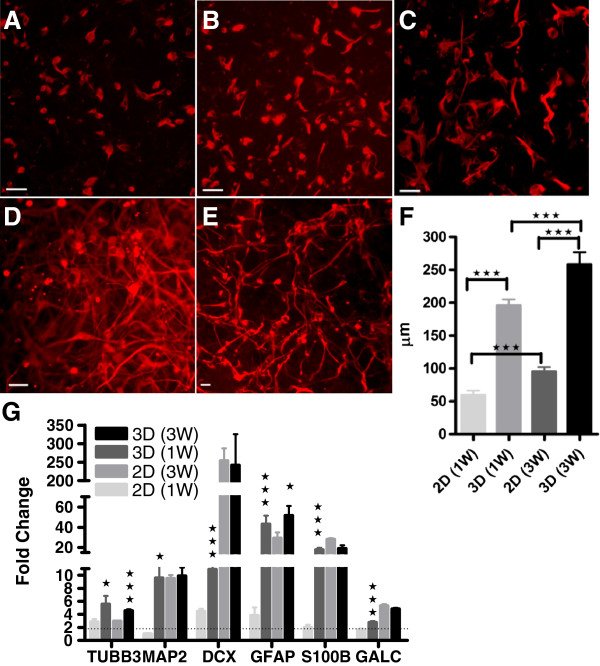
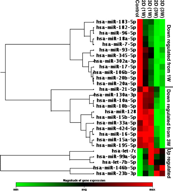
Methods: HNSCs were differentiated on 2- (2D) and 3-dimensional (3D) cultures for 1 and 3 weeks. Quantification of hNSC differentiation was measured with real-time PCR and axon outgrowth. The miRNA PCR arrays were implemented to investigate differential expression profiles in differentiated hNSCs. Evaluation of miRNA effects on hNSCs was performed by using transfection of miRNA mimics, real-time PCR, Western blot, and immunocytochemistry.

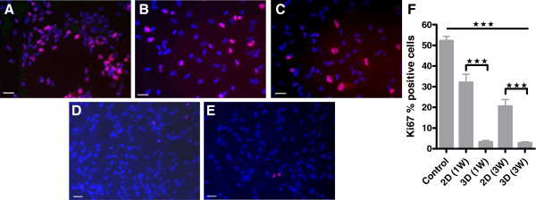
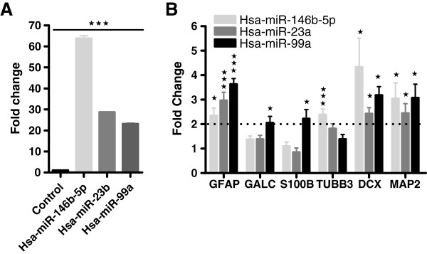
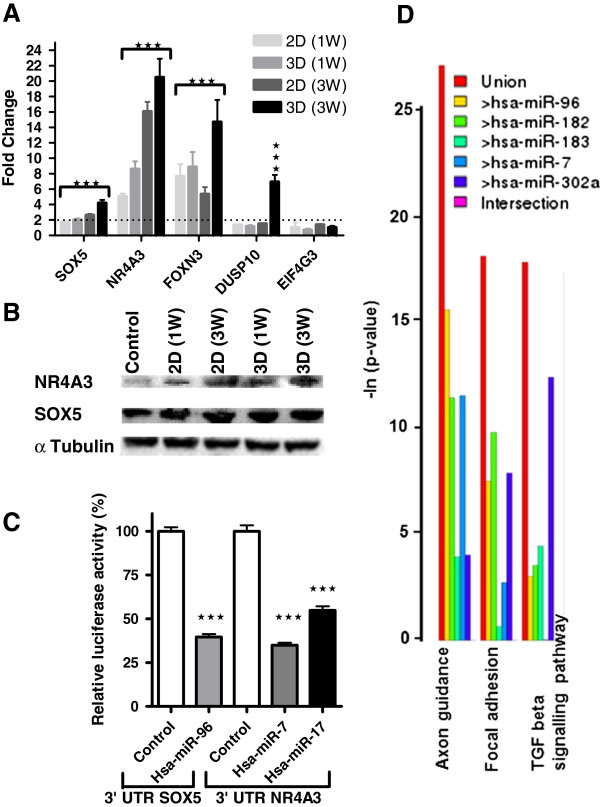
Results: The 3D substrate promoted enhanced hNSC differentiation coupled with a loss of cell proliferation. Differentiated hNSCs exhibited a similar miRNA profiling. However, in 3D samples, the degree and timing of regulation were significantly different in miRNA members of cluster mi-R17 and miR-96-182, and hsa-miR-302a. Overall, hNSC 3D cultures demonstrated differential regulation of miRNAs involved in hNSC stemness, cell proliferation, and differentiation. The miRNA mimic analysis of hsa-miR-146b-5p and hsa-miR-99a confirmed induction of lineage-committed progenitors. Downregulated miRNAs were more abundant; those most significantly downregulated were selected, and their putative target mRNAs analyzed with the aim of unraveling their functionality. In differentiated hNSCs, downregulated hsa-miR-96 correlated with SOX5 upregulation of gene and protein expression; similar results were obtained for hsa-miR-302a, hsa-miR-182, hsa-miR-7, hsa-miR-20a/b, and hsa-miR-17 and their target NR4A3. Moreover, SOX5 was identified as a direct target gene of hsa-miR-96, and NR43A, a direct target of hsa-miR-7 and hsa-mir-17 by luciferase reporter assays. Therefore, the regulatory role of these miRNAs may occur through targeting NR4A3 and SOX5, both reported as modulators of cell-cycle progression and axon length.

Conclusions: The results provide new insight into the identification of specific miRNAs implicated in hNSC differentiation. These strategies may be exploited to optimize in vitro hNSC differentiation potential for use in preclinical studies and future clinical applications.


Figures

Figure 1
Figure 1
Quantification of hNSC differentiation. (A-E) Representative image of (A) undifferentiated, (B) 1 week (1W) 2D differentiated, (C) 3-week (3W) 2D differentiated, (D) 1W 3D differentiated, and (E) 3W 3D differentiated hNSCs; scale bar, 50 μm. (F) Quantification of axon length on 1W and 3W differentiated hNSCs cultured on 2D and 3D substrates. (G) QRT-PCR molecular analysis for neuronal (TUBB3, DCX, and MAP2) and glial (GALC, GFAP, and S100B) markers performed on hNSCs differentiated on 2D and 3D for 1W, and 3W, and expressed as fold change compared with undifferentiated control. Statistical analysis showed significant differences between 2D versus 3D samples at the same time point; ± SDMs. *P < 0.05; **P < 0.001, ***P < 0.005, Student t test.
Figure 2
Figure 2
Evaluation of hNSC proliferation. (A-E) Representative image of (A) undifferentiated, (B) 1W 2D differentiated, (C) 3W 2D differentiated, (D) 1W 3D differentiated, and (E) 3W 3D differentiated hNSCs stained with Ki67, marker of cell proliferation; scale bar, 50 μm; Ki67+ hNSCs (red), nuclear Hoechst counterstain (blue). (F) Quantification of cell proliferation measured as percentage of cells positive for Ki67. Statistical analysis showed a significant reduction in proliferation in 2D and 3D cultures compared with the proliferative control and between 2D and 3D samples at the same time point; ±SDMs. *P < 0.05, **P < 0.001, ***P < 0.005, Student t test.
Figure 3
Figure 3
Human cell differentiation and development miScript miRNA PCR array profiles. Group clustergram analysis of miRNA PCR array profiles analysis of differentially regulated miRNAs identified in control (undifferentiated hNSCs) and hNSCs seeded on 2D and 3D substrates and differentiated for 1W and 3W.
Figure 4
Figure 4
MiRNA mimic transfection analysis. A) hsa-miR-146b-5p, hsa-miR-23b, hsa-miR-99a transfected mimics were measured by real-time RT-PCR, and expressed as fold change compared with GFP transfected hNSCs (control). B) QRT-PCR molecular analysis for neuronal (TUBB3, DCX, and MAP2) and glial (GALC, GFAP, and S100B) markers of 146b-5p, hsa-miR-23b, hsa-miR-99a mimic transfected hNSCs and expressed as fold change compared with GFP transfected hNSCs (control). Statistical analysis was performed against transfected control; ± SDMs, *p < 0.05, **p < 0.001, ***p < 0.005, Student’s t-test.
Figure 5
Figure 5
Validation of miRNA target-predicted genes. (A) qRT-PCR analysis performed on 1W and 3W differentiated hNSCs cultured on 2D and 3D substrates and expressed as fold change compared with proliferative control. Statistical analysis showed significant difference compared with control; ±SDMs, *P < 0.05, **P < 0.001, ***P < 0.005, Student t test. (B) SOX5 and NR4A3 protein quantification performed by Western blot on 1W and 3W differentiated hNSCs cultured on both 2D or 3D substrates and proliferative control. (C) Dual luciferase report assay. Measurement of the relative luciferase activity of SOX5 and NR4A3 3′-UTR constructs transfected with hsa-miR-96, and hsa-miR-7 and 17, respectively. Data are expressed as mean values ± SDMs and are shown as percentage of control (cells transfected with either SOX5 or NR4A3 3′-UTR constructs and control microRNA). Each bar represents values from three independent experiments, measured in triplicate. The relative activity of firefly luciferase expression was normalized to renilla luciferase activity. Data were analyzed with Student t test, ***P < 0.005. (D) MiRNA KEGG pathway analysis results obtained by using DIANA Lab.

References

    1. Falanga V. Stem cells in tissue repair and regeneration. J Invest Dermatol. 2012;132:1538–1541. doi: 10.1038/jid.2012.77. - DOI - PMC - PubMed
    1. Pollock K, Stroemer P, Patel S, Stevanato L, Hope A, Miljan E, Dong Z, Hodges H, Price J, Sinden JD. A conditionally immortal clonal stem cell line from human cortical neuroepithelium for the treatment of ischemic stroke. Exp Neurol. 2006;199:143–155. doi: 10.1016/j.expneurol.2005.12.011. - DOI - PubMed
    1. Hodges H, Pollock K, Stroemer P, Patel S, Stevanato L, Reuter I, Sinden J. Making stem cell lines suitable for transplantation. Cell Transplant. 2007;16:101–115. - PubMed
    1. Smith EJ, Stroemer RP, Gorenkova N, Nakajima M, Crum WR, Tang E, Stevanato L, Sinden JD, Modo M. Implantation site and lesion topology determine efficacy of a human neural stem cell line in a rat model of chronic stroke. Stem Cells. 2012;30:785–796. doi: 10.1002/stem.1024. - DOI - PubMed
    1. Stroemer P, Patel S, Hope A, Oliveira C, Pollock K, Sinden J. The neural stem cell line CTX0E03 promotes behavioral recovery and endogenous neurogenesis after experimental stroke in a dose-dependent fashion. Neurorehabil Neural Repair. 2009;23:895–909. doi: 10.1177/1545968309335978. - DOI - PubMed

Publication types

Associated data