. 2014 May 8;54(3):335-48.
doi: 10.1016/j.molcel.2014.03.018. Epub 2014 Apr 10.

Molecular basis and regulation of OTULIN-LUBAC interaction

Affiliations

Molecular basis and regulation of OTULIN-LUBAC interaction

Paul R Elliott et al. Mol Cell. .

Abstract

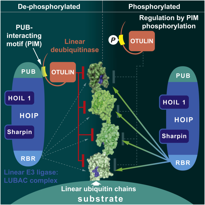
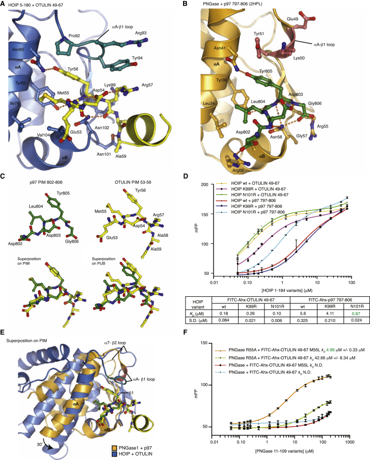
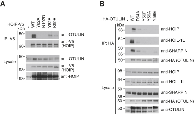
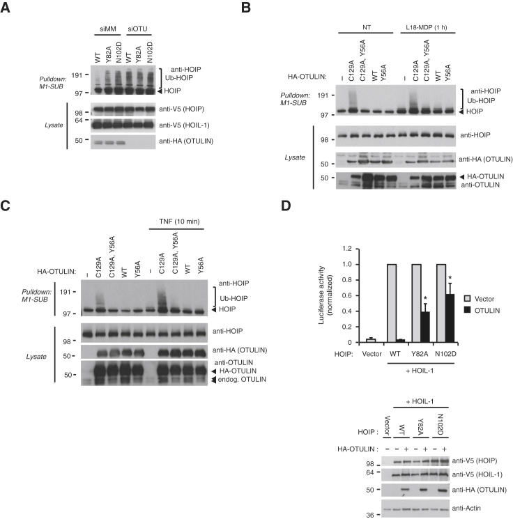
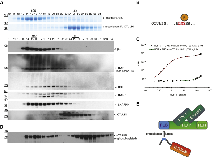
The linear ubiquitin (Ub) chain assembly complex (LUBAC) generates Met1-linked "linear" Ub chains that regulate the activation of the nuclear factor κB (NFκB) transcription factor and other processes. We recently discovered OTULIN as a deubiquitinase that specifically cleaves Met1-linked polyUb. Now, we show that OTULIN binds via a conserved PUB-interacting motif (PIM) to the PUB domain of the LUBAC component HOIP. Crystal structures and nuclear magnetic resonance experiments reveal the molecular basis for the high-affinity interaction and explain why OTULIN binds the HOIP PUB domain specifically. Analysis of LUBAC-induced NFκB signaling suggests that OTULIN needs to be present on LUBAC in order to restrict Met1-polyUb signaling. Moreover, LUBAC-OTULIN complex formation is regulated by OTULIN phosphorylation in the PIM. Phosphorylation of OTULIN prevents HOIP binding, whereas unphosphorylated OTULIN is part of the endogenous LUBAC complex. Our work exemplifies how coordination of ubiquitin assembly and disassembly activities in protein complexes regulates individual Ub linkage types.


Figures

None
Graphical abstract
Figure 1
Figure 1
OTULIN Binds the HOIP PUB Domain (A) Epitope-tagged HOIP or HOIL-1 were transfected into HEK293T cells, and interaction with endogenous OTULIN was determined by immunoprecipitation followed by western blot analysis. OTULIN interacts with HOIP but not HOIL-1 under these conditions. (B) Domain representation of HOIP. A bar graph below indicates constructs used for domain mapping. (C) Domains of epitope-tagged HOIP were transfected into U2OS and NOD2 cells and probed for endogenous OTULIN following the coimmunoprecipitation described in (A). (D) Analytical size-exclusion chromatography profile of HOIP 1-184 (blue), full-length OTULIN (red), and 1:1.2 OTULIN:HOIP complex (black). Coomassie-stained SDS-PAGE gels below show protein-containing fractions. (E) Left, extended HOIP PUB domain structure (blue). Middle left, HOIP PUB domain structure determined by the SGC (green, PDB ID 4JUY). Middle right, structure of PNGase PUB domain (orange, PDB ID 2HPL) (Zhao et al., 2007). Right, superposition. The SGC-determined HOIP structure includes an additional TEV protease cleavage site at the N terminus (see also Figure S5B). (F) Structure-based sequence alignment of HOIP and PNGase PUB domains. HOIP contains two additional N-terminal helices and an additional C-terminal helix not found in PNGase. Open circles represent residues in HOIP (blue), and PNGase (yellow) that interact with the OTULIN/p97 PIMs, respectively.
Figure 2
Figure 2
A PUB-Domain-Interacting Motif in OTULIN (A) Structure of PNGase bound to the p97 PIM peptide (PDB ID 2HPL) (Zhao et al., 2007) reveals the position of the PIM pocket. (B) Surface conservation analysis of the HOIP PUB domain colored according to the sequence alignment in Figure S1A. The PIM pocket is highly conserved, whereas other regions, including the surface generated by the N-terminal PUB domain extension, are not conserved. (C) Primary sequence alignment of the HOIP binding region in OTULIN (Figure S3A) (Rivkin et al., 2013). Alignment shows that the patch with highest evolutionary conservation resembles the p97 PIM. (D) Affinity measurements using HOIP PUB domain against FITC-Ahx-labeled p97 PIM peptide (aa 797–806) and OTULIN PIM peptide (aa 49–67). Experiments were performed in triplicate, and errors represent SD from the mean. (E) Binding of PUB domains from HOIP (aa 1–184, red), PNGase (aa 11–109, green), and UBXD1 (aa 150–264, magenta) to a fluorescent p97 PIM peptide (aa 797–806) as in (D). KD values are indicated, and errors represent SD from the mean from triplicate experiments. (F) Binding of PUB domains as in (E) to the OTULIN PIM peptide. (G) 15N-transverse relaxation optimized spectroscopy (TROSY) spectra of HOIP alone (black), HOIP bound to OTULIN PIM peptide at a 1:1 molar ratio (red), and HOIP bound to p97 PIM peptide at a 1:1 (blue) and 1:4 ratio (green). Selected perturbed resonances are shown. For the full spectra, see Figure S4A. (H) Chemical shift map by HOIP residue number for perturbation by p97 and OTULIN PIM peptides. (I) 15N-TROSY spectra of HOIP alone (black), HOIP bound to OTULIN PIM peptide (red), HOIP bound to full-length OTULIN (blue), and HOIP with OTULIN catalytic domain (aa 80–352, yellow), all at a 1:1 molar ratio. The same resonances as in (G) are shown. For the full spectra, see Figures S4B, 4C, and 3. (J) Difference map of chemical shifts between HOIP bound to PIM peptide or full-length OTULIN derived from respective spectra in (I).
Figure 3
Figure 3
Structure of HOIP Bound to OTULIN Peptide (A) Structure of HOIP PUB domain (aa 5–180; blue) bound to the OTULIN PIM peptide (yellow). The peptide is in ball-and-stick representation with blue nitrogen and red oxygen atoms. (B) A weighted 2|Fo|-|Fc| map contoured at 1 σ covering the OTULIN PIM peptide colored as in (A). (C) LIGPLOT representation of the HOIP-OTULIN interaction. Residues in the PIM (aa 53–57) are shown in yellow, and the C-terminal extension of the PIM is shown in orange. Hydrogen bonds are shown by green dashes, and van der Waals contacts are shown as red fans. (D) PIM pocket shown in surface representation in the HOIP-OTULIN complex colored as in (A). Residues 92–94, including the mobile Tyr94, are colored green. (E) Close-up view of the OTULIN PIM peptide in the HOIP PIM pocket, colored as in (A), showing hydrogen bonds as orange dotted lines. (F) PIM pocket shown in surface representation in apo HOIP, in which Tyr94 (green) partly occludes the PIM pocket. (G) Superposition of apo and PIM-peptide-bound HOIP highlighting the conformational change in Tyr94 side chain. (H) A conformational change of the Tyr94 side chain is resolved in the aromatic region of 13C-HSQC spectra, with HOIP alone (black) and the HOIP OTULIN PIM complex spectrum shown in red. The shifting resonance indicated by an arrow corresponds to the Cε of Tyr94. Only the Cε region of the aromatic 13C-HSQC is shown. (I) Fluorescent polarization assay of wild-type OTULIN PIM peptide binding to purified HOIP (aa 1–184) PIM pocket mutants. Binding parameters are listed below. Experiments were performed in triplicate, and errors represent SD from the mean. (J) Binding of HOIP PUB domain (aa 1–184) to OTULIN peptides (aa 49–67) with the indicated point mutations in the PIM performed as in (I). Binding parameters are listed below.
Figure 4
Figure 4
Specificity of the HOIP-OTULIN Interaction (A) Close-up view of the HOIP PUB domain (blue) bound to OTULIN PIM (yellow) as in Figure 3E. Interacting residues are shown in ball-and-stick representation and labeled. Hydrogen bonds are indicated as orange dotted lines. (B) Same view as in (A) for the PNGase-p97 complex (PDB ID 2HPL) (Zhao et al., 2007). Residues 49–51 that differ structurally from HOIP are colored red. (C) PIM peptides from p97 (green) and OTULIN (yellow) can be perfectly superimposed (bottom left) but do not align once PUB domains are superposed because of deeper binding of the OTULIN PIM in the HOIP PIM pocket. (D) Fluorescence polarization assays of HOIP N101R/K99R mutants with FITC-Ahx-labeled p97 (797–806) or OTULIN (49–67) PIMs as described in Figure 2D. Binding parameters are listed below. Experiments were performed in triplicate, and errors represent SD from the mean. (E) Superposition on the PIM of PNGase-p97 (orange and green) and HOIP-OTULIN (blue and yellow) shows perfect alignment of the Asn cornerstone residue but also the misalignment of PUB domain core helices, indicating different binding modes. (F) Fluorescence polarization assays of PNGase and FITC-Ahx-labeled OTULIN (49–67) with point mutations in the PUB domain and the PIM peptide that promote binding of OTULIN PIM to PNGase. Experiments were performed in triplicate, and errors represent SD from the mean.
Figure 5
Figure 5
Verification of HOIP-OTULIN Interactions in Cells (A) Experiments performed as in Figure 1A with HOIP point mutations in the PIM pocket and testing the binding of endogenous OTULIN as detected by an OTULIN antibody. (B) HA-tagged OTULIN or OTULIN PIM mutants were expressed in HEK293T cells immunoprecipitated with anti-HA-Agarose resin, and LUBAC components HOIP, HOIL-1, and SHARPIN were detected by western blotting against endogenous components.
Figure 6
Figure 6
Functional Consequences of the OTULIN-LUBAC Interaction (A) Purification of endogenous Ub conjugates with Met1-specific Ub binding domain (Keusekotten et al., 2013) in lysates of HEK293T control and OTULIN-depleted cells transfected with HOIP variants and HOIL-1. Purified material and lysate was examined by immunoblotting. Mutation of the HOIP PIM pocket results in spontaneous accumulation of Met1-linked polyubiquitin on HOIP. (B and C) Purification of endogenous Ub conjugates with M1-SUB in U2OS and NOD2 cells transfected with the indicated OTULIN variants and treated with L18-MDP (B) or TNF (C). Purified material was analyzed as in (A). Mutation of the OTULIN PIM impairs stabilization of HOIP ubiquitination by catalytic inactive (C129A) OTULIN under basal conditions and after stimulation. (D) NFκB reporter activity in lysates of HEK293T cells transfected with HOIL-1, HOIP, or HOIP PIM pocket mutants and with or without the expression of OTULIN. OTULIN abrogated NFκB activity induced by wild-type LUBAC but was less effective in inhibiting activity induced by HOIP PIM pocket mutants.
Figure 7
Figure 7
Regulation of OTULIN-LUBAC Complex Formation by Phosphorylation (A) Gel filtration analysis of purified bacterial p97 hexamers and full-length OTULIN visualized by Coomassie staining and HEK293ET cell lysates probed with indicated antibodies. (B) Schematic of the OTULIN PIM indicating phosphorylation at Tyr56 as identified in 22 independent mass spectrometry experiments in http://phosphosite.org/proteinAction.do?id=2470471. (C) Fluorescence polarization assays of HOIP PUB domains with wild-type and Tyr56-phosphorylated FITC-Ahx-labeled OTULIN (49–67). Experiments were performed in triplicate, and errors represent SD from the mean. (D) HEK293ET lysates were prepared in absence of phosphatase inhibitors and probed for the same components as in (A). Only the OTULIN blot is shown. (E) Schematic model of the LUBAC-OTULIN complex indicating its regulation by protein phosphorylation.

Similar articles

Cited by

References

    1. Allen M.D., Buchberger A., Bycroft M. The PUB domain functions as a p97 binding module in human peptide N-glycanase. J. Biol. Chem. 2006;281:25502–25508. - PubMed
    1. Behrends C., Harper J.W. Constructing and decoding unconventional ubiquitin chains. Nat. Struct. Mol. Biol. 2011;18:520–528. - PubMed
    1. Boisson B., Laplantine E., Prando C., Giliani S., Israelsson E., Xu Z., Abhyankar A., Israël L., Trevejo-Nunez G., Bogunovic D. Immunodeficiency, autoinflammation and amylopectinosis in humans with inherited HOIL-1 and LUBAC deficiency. Nat. Immunol. 2012;13:1178–1186. - PMC - PubMed
    1. Chen Z.J., Sun L.J. Nonproteolytic functions of ubiquitin in cell signaling. Mol. Cell. 2009;33:275–286. - PubMed
    1. Emmerich C.H., Ordureau A., Strickson S., Arthur J.S.C., Pedrioli P.G.A., Komander D., Cohen P. Activation of the canonical IKK complex by K63/M1-linked hybrid ubiquitin chains. Proc. Natl. Acad. Sci. USA. 2013;110:15247–15252. - PMC - PubMed

Publication types

Associated data