Review
. 2014 Jul 1;391(1):1-16.
doi: 10.1016/j.ydbio.2014.04.001. Epub 2014 Apr 12.

Cell signaling during development of Dictyostelium

Affiliations
Review

Cell signaling during development of Dictyostelium

William F Loomis. Dev Biol. .

Abstract

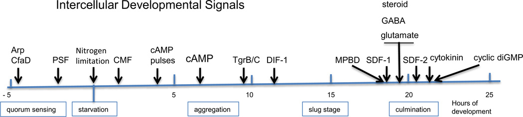
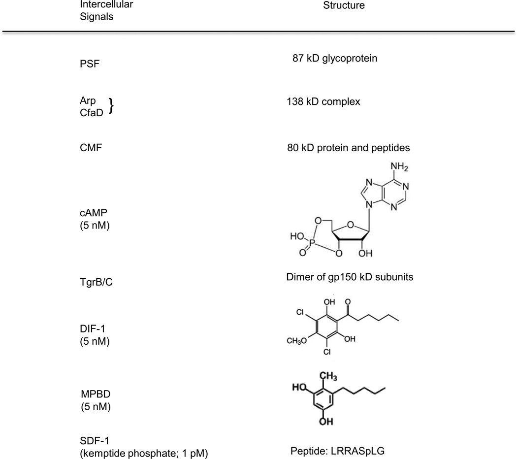
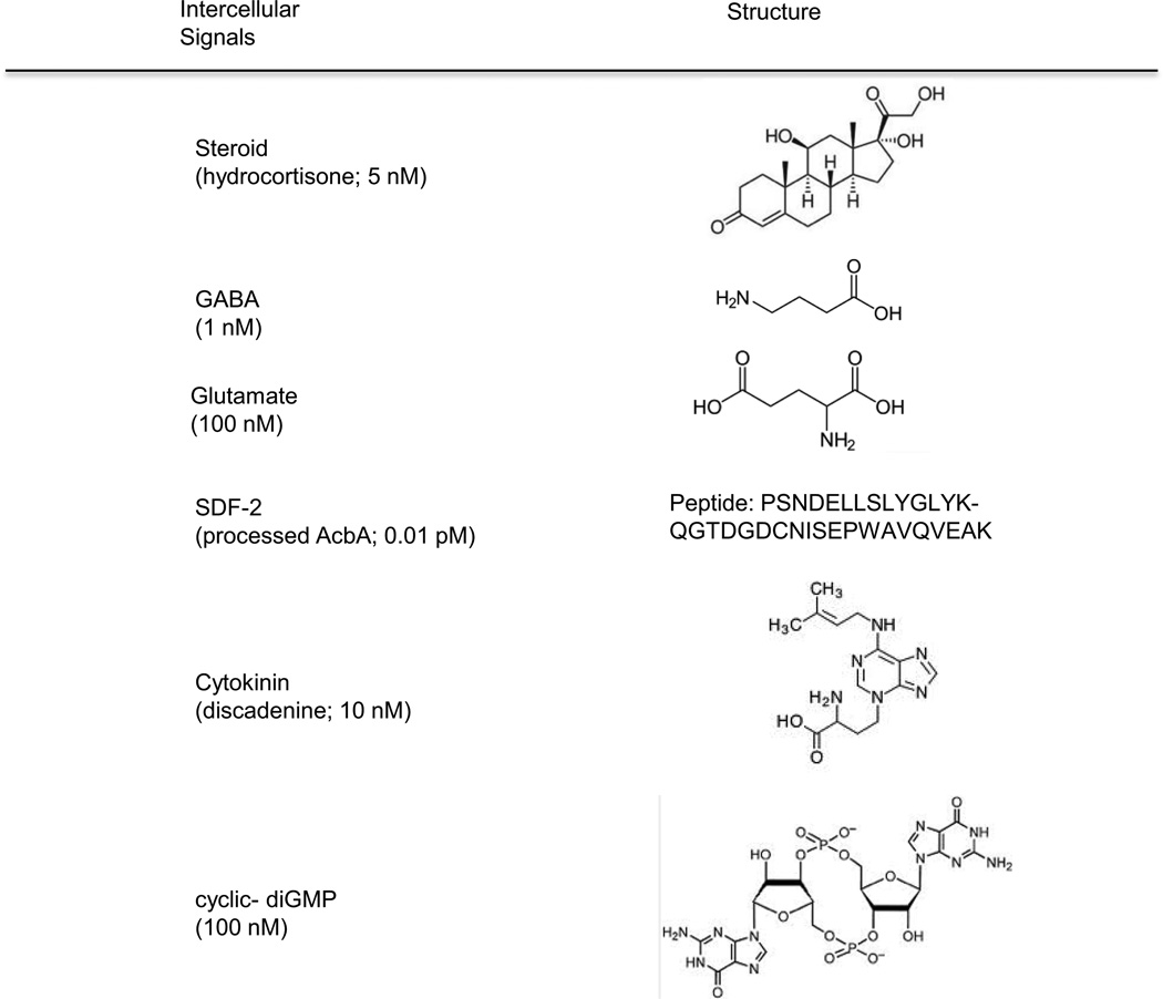
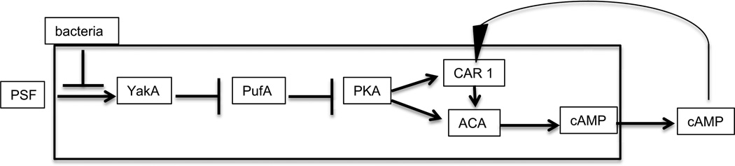
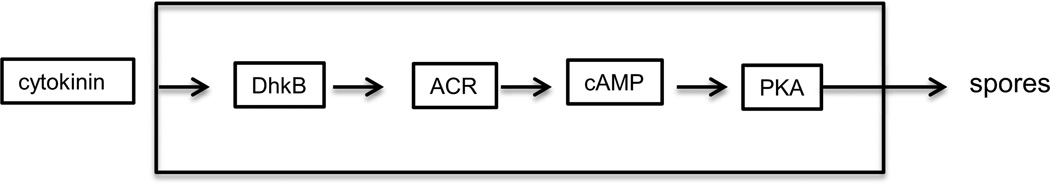
Continuous communication between cells is necessary for development of any multicellular organism and depends on the recognition of secreted signals. A wide range of molecules including proteins, peptides, amino acids, nucleic acids, steroids and polylketides are used as intercellular signals in plants and animals. They are also used for communication in the social ameba Dictyostelium discoideum when the solitary cells aggregate to form multicellular structures. Many of the signals are recognized by surface receptors that are seven-transmembrane proteins coupled to trimeric G proteins, which pass the signal on to components within the cytoplasm. Dictyostelium cells have to judge when sufficient cell density has been reached to warrant transition from growth to differentiation. They have to recognize when exogenous nutrients become limiting, and then synchronously initiate development. A few hours later they signal each other with pulses of cAMP that regulate gene expression as well as direct chemotactic aggregation. They then have to recognize kinship and only continue developing when they are surrounded by close kin. Thereafter, the cells diverge into two specialized cell types, prespore and prestalk cells, that continue to signal each other in complex ways to form well proportioned fruiting bodies. In this way they can proceed through the stages of a dependent sequence in an orderly manner without cells being left out or directed down the wrong path.

Keywords: Dependent sequence; Intercellular communication; Signal transduction.


Figures

Figure 1
Figure 1
Signaling during development. The signals used to integrate development of Dictyostelium are indicated at the stages at which they act. In the 5 hours preceeding the initiation of development while the cells are still growing secreted proteins function as quorum sensors. Morphogenesis occurs over 24 hours following the initiation of development by nitrogen limitation; the stages are indicated below the temporal line. The structure of each signal is given in Figure 2 and the mode of action described in the text.
Figure 2
Figure 2
Structures of the intercellular signals. The signals used by Dictyostelium for communication among developing cells are highly diverse and include proteins, peptides, polyketides, nucleic acids, amino acids and steroids. Each signal is recognized by a unique receptor on the surface that initiates a signal transduction pathway within the cells. Many of the small molecules are similar or identical to signals used by mammalian tissues. Others are similar to signals used by plants, yeast or bacteria.
Figure 2
Figure 2
Structures of the intercellular signals. The signals used by Dictyostelium for communication among developing cells are highly diverse and include proteins, peptides, polyketides, nucleic acids, amino acids and steroids. Each signal is recognized by a unique receptor on the surface that initiates a signal transduction pathway within the cells. Many of the small molecules are similar or identical to signals used by mammalian tissues. Others are similar to signals used by plants, yeast or bacteria.
Figure 3
Figure 3
The growth- differentiation transition pathway. Growing amoebae secrete PSF continuously such that the concentration increases as the cell density increases. When it reaches threshold, it activates the protein kinase YakA. If bacteria are still around, the threshold is higher. YakA activity inhibits PufA which inhibits translation of the catalytic subunit of PKA. Inhibiting an inhibitor results in activation of PKA. This cAMP dependent protein kinase leads to the accumulation of the cAMP receptor CAR1 and the adenylyl cyclase ACA which synthesizes cAMP. Most of the newly made cAMP is secreted into the surrounding fluid where it can diffuses to bind to the receptor on the same cell (autocrine) or other cells (paracrine). Ligand binding to CAR1 stimulates ACA thereby forming a positive feedback loop.
Figure 4
Figure 4
Quorum sensing. Growing amoebae secrete two proteins, AprA and CfaD, that act as quorum sensors and limit cell proliferation before exogenous nutrients have been fully used up, giving the cells a little extra time. A G protein coupled receptor (GPCR) is implicated by the requirement for the G protein subunit Ga8. The DNA binding protein BzpN inhibits proliferation predominantly at low cell density and the protein kinase PakD inhibits proliferation predominantly at high cell density. PakD is also essential for developmental aggregation.
Figure 5
Figure 5
Modulation of cAMP signaling and control of developmental genes. The 80 kDa protein CMF affects early development in at least two independent manners. Shortly after the initiation of development CMF is secreted and signals whether there is a sufficient density of cells to make it worthwhile to aggregate and form fruiting bodies. If CMF is present at 10 ng/ml or higher, G proteins activated by CAR1 are able to stimulate the adenylyl cyclase ACA to synthesize cAMP. Most of the newly made cAMP is secreted so that it can bind to the CAR1 receptor thereby closing a positive feedback loop. CMF and small peptides that are cleaved from it can bind to the receptor CMFR1 which leads to a signal transduction pathway ending in expression of the marker genes cotB and cprD.
Figure 6
Figure 6
The PKA oscilatory circuit. Binding of cAMP to its receptor CAR1 not only activates ACA through its trimeric G protein but also activates the MAP kinase Erk2. This protein kinase inhibits the internal cAMP phosphodiesterase RegA such that it no longer reduces the internal level of cAMP. Increasing the rate of synthesis of cAMP and decreasing its rate of degradation leads to a surge in the concentration of cAMP. Most of the newly synthesized cAMP is secreted where it can further stimulate the circuit. However, internal cAMP activates the protein kinase PKA. Acting indirectly, PKA leads to a block in the activation of ACA and also activates the transcription factor GataC. The reduction in ACA activity lowers the levels of cAMP and the circuit proceeds to reset. External cAMP is reduced by the secreted cAMP phosphodiesterase PdsA which interfers with stimulation of the circuit. Although phosphorylation activates GataC, it also leads to its exit from the nucleus. As a result, GataC is able to stimulate transcription of developmental genes only for a brief period following each pulse of cAMP. The pulse induced genes are still expressed in mutant strains lacking ACA as the result of sufficient cAMP being synthesized by the minor adenylyl cyclase ACR to activate PKA when RegA is inhibited by Erk2.
Figure 7
Figure 7
Control of ACA dependent genes. A set of at least 13 developmental genes are only expressed if the gene for ACA is wild type. The signal transduction pathway from the extracellular cAMP signal to transcriptional control is almost the same as that in the PKA oscilatory circuit (Fig. 6) but requires the robust synthesis of cAMP that ACA can provide following a pulse of extracellular cAMP. It is possible that transcription of these genes depends on higher levels of PKA than the pulse induces genes.
Figure 8
Figure 8
Cell contact signaling. A feedforward loop controls the expression of GBF dependent genes. GBF is a DNA binding protein that regulates expression of many developmental genes including the tgr genes that encode transmembrane proteins for cell-cell adhesion. The Tgr proteins are highly polymorphic in nature and act in intercellular signaling to indicate the level of kinship. If TrgB and TgrC are compatible in adjacent cells, then the GBF dependent developmental genes are expressed as long as GBF is also functional. Such a feedforward loop acts as a low pass filter to avoid reacting to short lived fluctuations in the signals.
Figure 9
Figure 9
Control of prestalk genes by DIF. The chlorinated hexaphenone DIF-1 is synthesized by prespore cells and degraded by prestalk cells. It induces PstO genes via the heterodimer of DNA binding proteins DimA/DimB. Not all genes expressed in PstO cells depend on DIF-1 indicating that it acts in a gene specific manner and not just as a general inducer of PstO differentiation.
Figure 10
Figure 10
SDF-1 signaling. The small phosphopeptide SDF-1 is secreted after 18 hours of development and signals both prestalk and prespore to prepare themselves for terminal differentiation. This process requires both RNA and protein synthesis. SDF-1 is cleaved from a larger phosphoprotein that is secreted in response to signaling by a polyketide, MPBD, or an increase in the activity of PKA. MPBD is synthesized by the Steely protein, StlA, and binds to the GPCR receptor CrlA. The signal is transduced by the trimeric protein that contains the Gα1 subunit such that the protein kinase GskA is inhibited and no longer blocks release of the SDF-1 precursor protein. The precursor is processed into SDF-1 by the extracellular protease domain of the prestalk specific protein TagB. SDF-1 stimulates PKA activity in a process that depends on the adenylyl cyclase ACG. High PKA activity can also trigger release of the SDF-1 precursor protein such that low levels of SDF-1 are amplified by "priming'.
Figure 11
Figure 11
SDF-2 signaling. A cascade of intercellular signals amplifies the first signal leading to extracellular production of SDF-2 which is mediated by a steroid that is very similar to hydrocortisone. The steroid receptor is a surface GPCR coupled to a trimeric G protein with the Ga4 subunit. This pathway leads to the release of GABA. The GABA receptor is a surface GPCR coupled to trimeric G protein with the Ga7 subunit. This same receptor binds glutamate when it is coupled to the trimeric G protein with the Ga9 subunit. Glutamate binding inhibits release of the SDF-1 precursor acyl-coA-binding protein, AcbA, but has a lower affinity to GrlE than GABA. When GABA accumulates in the intercellular space the lipid kinase PI3K and the protein kinase PKB R1 are activated and release of AcbA is stimulated. AcbA is cleaved by the extracellular protease domain of the prestalk specific protein TagC to generate SDF-2. This 34 amino acid peptide has to diffuse back to the prespore cells where it triggers rapid encapsulation into spores. The SDF-2 receptor is the membrane embedded histidine kinase DhkA that is converted to a protein phosphatase when SDF-2 is bound. It then removes the phosphate from the small H2 RdeA so that it can no longer phosphorylate and activate the internal cAMP phosphodiesterase RegA. In fact, it actively dephosphorylates RegA thereby inhibiting it from degrading cAMP. As cAMP builds up, PKA is activated and can trigger spore formation as well as release of further AcbA.
Figure 12
Figure 12
Cytokinin signaling. Cytokinins are thought of as plant hormones but their use as intercellular signals in Dictyostelium results from inheritance from a common ancestor of amoebozoa and plants. The isopentyladenine derivative discadenine is a cytokinin produced by Dictyostelium that leads to an increase in PKA activity in a manner dependent on the histidine kinase, DhkB and the late adenylyl cyclase ACR. As is the case in SDF-2 signaling, the increase in PKA activity leads to rapid encapsulation of prespore cells. DhkB and ACR are preferentially found in prespore cells.

References

    1. Abe H, Uchiyama M, Tanaka Y, Saito H. Structure of discadenine, a spore germination inhibitor from the cellular slime mold Dictyostelium discoideum. Tetrahedron Lett. 1976;42:3807–3810.
    1. Anjard C, Loomis WF. Peptide signaling during terminal differentiation of Dictyostelium. Proc. Natl. Acad. Sci. USA. 2005;102:7607–7611. - PMC - PubMed
    1. Anjard C, Loomis WF. GABA induces terminal differentiation of Dictyostelium through a GABA(B) receptor. Development. 2006;133:2253–2261. - PubMed
    1. Anjard C, Loomis WF. Cytokinins induce sporulation in Dictyostelium. Development. 2008;135:819–827. - PubMed
    1. Anjard C, Su Y, Loomis WF. Steroids initiate a signaling cascade that triggers rapid sporulation in Dictyostelium. Development. 2009;136:803–812. - PMC - PubMed

Publication types

LinkOut - more resources