Review
. 2014 Jul:72:23-40.
doi: 10.1016/j.freeradbiomed.2014.03.039. Epub 2014 Apr 12.

Physiology and pathophysiology of iron in hemoglobin-associated diseases

Affiliations
Review

Physiology and pathophysiology of iron in hemoglobin-associated diseases

Thomas D Coates. Free Radic Biol Med. 2014 Jul.

Abstract

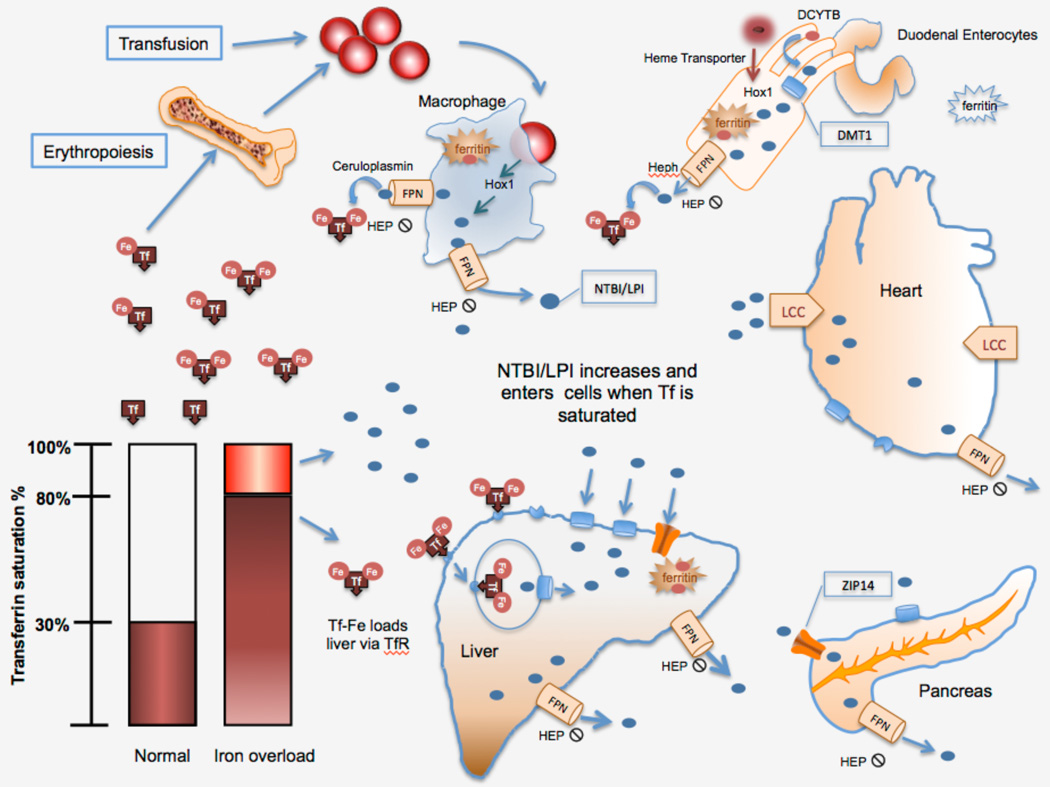
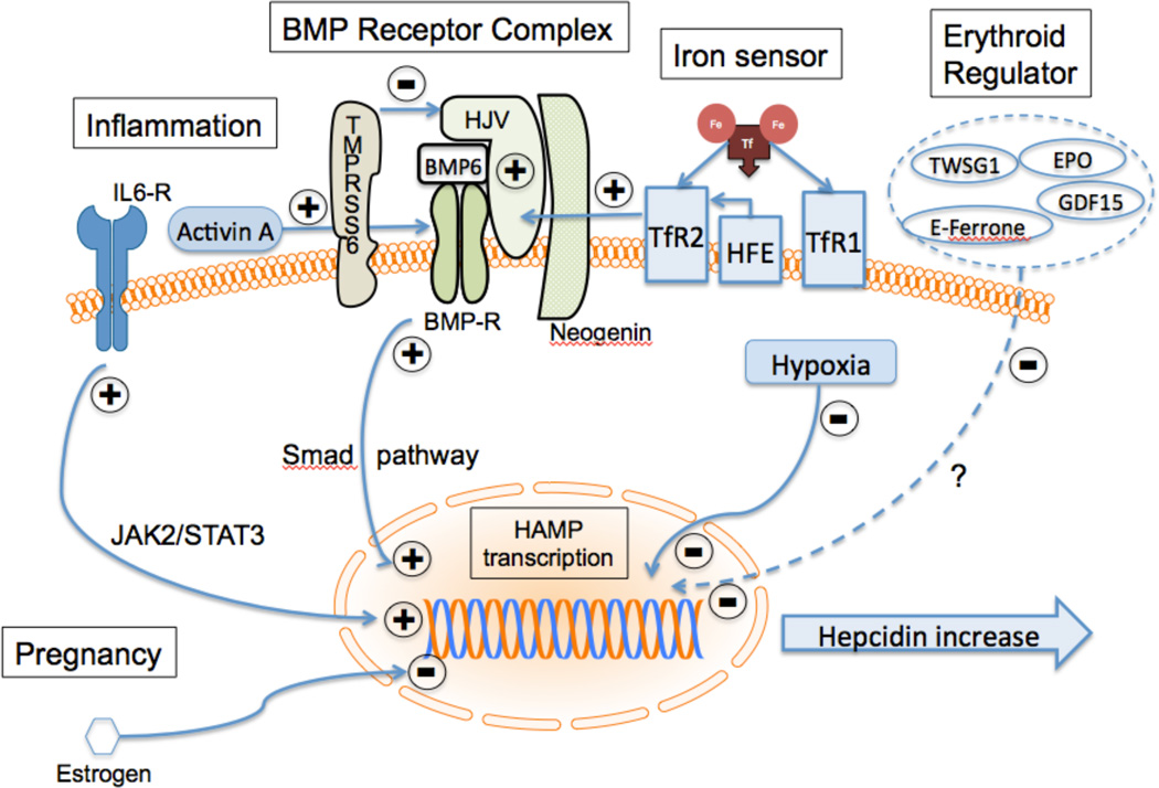
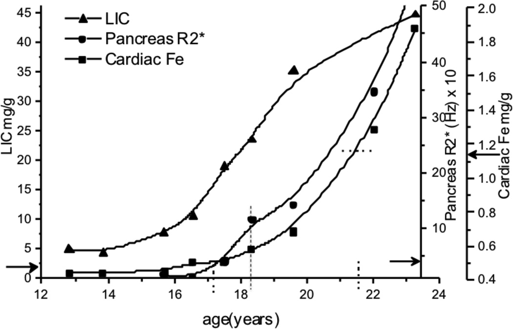
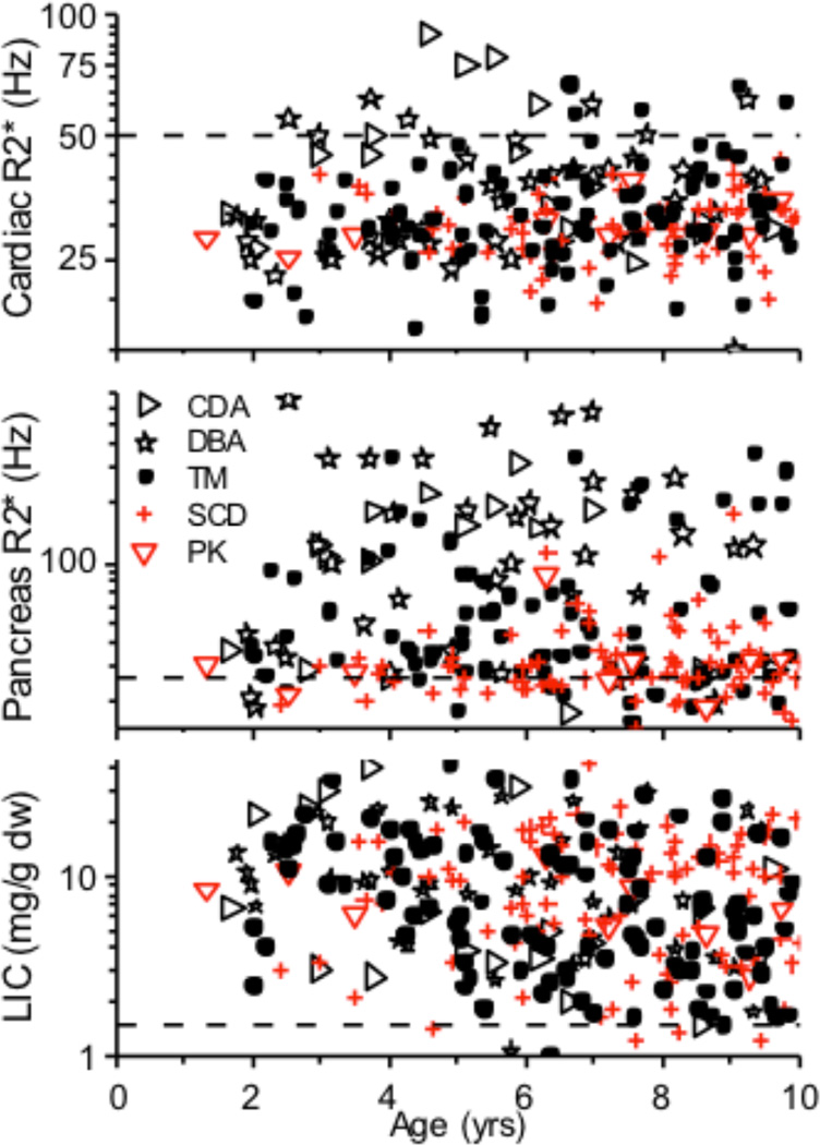
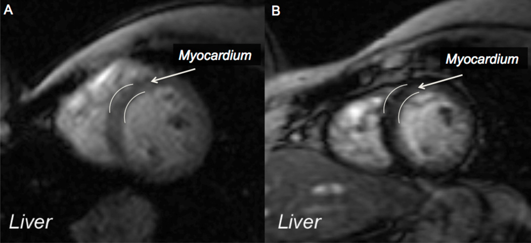
Iron overload and iron toxicity, whether because of increased absorption or iron loading from repeated transfusions, can be major causes of morbidity and mortality in a number of chronic anemias. Significant advances have been made in our understanding of iron homeostasis over the past decade. At the same time, advances in magnetic resonance imaging have allowed clinicians to monitor and quantify iron concentrations noninvasively in specific organs. Furthermore, effective iron chelators are now available, including preparations that can be taken orally. This has resulted in substantial improvement in mortality and morbidity for patients with severe chronic iron overload. This paper reviews the key points of iron homeostasis and attempts to place clinical observations in patients with transfusional iron overload in context with the current understanding of iron homeostasis in humans.

Keywords: Chelation; Hemochromatosis; Hemoglobinopathy; Iron overload; Iron toxicity; Magnetic resonance imaging; ROS; Sickle cell disease; Thalassemia; Transfusion.


Conflict of interest statement

Conflict of interest: Dr Coates is on the Speaker’s Bureau or consults for Novartis Pharma, Shire Pharma, Apo Pharma, and Celeron.

Figures

Figure 1
Figure 1
Iron homeostasis in transfusional iron overload. Red cells (RBC), phagocytosed by reticuloendothelial macrophages, the hemoglobin is degraded by heme oxygenase (HOX-1) and Fe is exported via ferroportin (FPN) and binds to transferrin (Tf). When Tf becomes saturated, non-transferrin bound iron (NTBI) and labile plasma iron (LPI) can enter organs through the divalent metal transported (DMT1), ZIP14, and L-type calcium channels (LCC). LPI and labile cellular iron (LCI) are highly reactive species of NTBI that are able to cause direct oxidant damage. Diferric transferrin enters the marrow and liver through the transferrin receptors 1 & 2. Heme and ferric iron enter the gut and are exported by FPN. Hepcidin (HEP) blocks export of Fe through FPN.
Figure 2
Figure 2
Regulation of hepcidin production. Bone morphogenetic protein-6 (BMP6) activates transcription of the hepcidin gene (hepcidin antimicrobial peptide; HAMP) via the SMAD pathway. Hemojuvelin (HJV) enhances the activity of BMP receptor (BMP-R) and this activity is suppressed by cleavage of HJV by TMPRSS6. Diferric Tf displaces the hemochromatosis protein (HFE) from the high affinity transferrin receptor, TfR1. It associates with TfR2 and this complex enhances signalling via BMP-R. In response to inflammation, activin-A can enhance BMP-R signaling or interleukin-6 acting through it receptor (IL6-R) can activate HAMP transcription. Twisted gastrulation-1 (TWSG1), grown differentiation factor 15 (GDF-15), erythroferrone (E-ferrone), estrogen, erythropoietin (EPO) and hypoxia reduce HAMP transcription.
Figure 3
Figure 3
Sequence of iron loading secondary to transfusion in the liver (LIC), pancreas, and heart in a single hemoglobinopathy patient. Solid arrows on the Y-axes mark upper normal levels. Pancreas begins to load at age 17 (vertical dotted line) and reaches very high levels by age 18.2 years. Cardiac loading does not reach clinically significant levels until age 21.5 yrs.
Figure 4
Figure 4
Iron loading (log scale) in transfused children with congenital dyserythropoietic anemia (CDA), Diamond-Blackfan anemia (DBA), pyruvate kinase deficiency (PK), sickle cell disease (SCD) and thalassemia major (TM). Children with ineffective erythropoiesis (black symbols) and those with effective (red symbols) erythropoiesis have similar iron loading of the liver at an early age. Children with ineffective or markedly decreased erythropoiesis (TM, CDA, DBA) have comparatively more loading of their pancreas and heart, consistent with NTBI-mediated loading. Dashed lines indicate upper limit of normal ranges.
Figure 5
Figure 5
Magnetic resonance images (MRI) of the chest showing very black liver indicating high iron and grey left ventricular wall indicating little iron (A). Two years later (B), the myocardium had loaded significantly and is black. The patient had become compliant with his chelation when he learned his heart was loaded with iron. The liver was cleared by chelation, but the heart was not. (Images courtesy of Dr. John Wood)

Similar articles

Cited by

References

    1. Berdoukas V, Farmaki K, Carson S, Wood J, Coates T. Treating thalassemia major-related iron overload: the role of deferiprone. Journal of blood medicine. 2012;3:119–129. - PMC - PubMed
    1. Modell B, Khan M, Darlison M, Westwood MA, Ingram D, Pennell DJ. Improved survival of thalassaemia major in the UK and relation to T2* cardiovascular magnetic resonance. J Cardiovasc.Magn Reson. 2008;10:42. - PMC - PubMed
    1. Green R, Charlton R, Seftel H, Bothwell T, Mayet F, Adams B, Finch C, Layrisse M. Body iron excretion in man: a collaborative study. Am J Med. 1968;45:336–353. - PubMed
    1. Ganz T. Systemic iron homeostasis. Physiol Rev. 2013;93:1721–1741. - PubMed
    1. Frazer DM, Anderson GJ. The regulation of iron transport. BioFactors. 2013 - PubMed

MeSH terms