. 2014 May 23;114(11):1690-9.
doi: 10.1161/CIRCRESAHA.1134303145. Epub 2014 Apr 11.

Fli1 acts downstream of Etv2 to govern cell survival and vascular homeostasis via positive autoregulation

Affiliations

Fli1 acts downstream of Etv2 to govern cell survival and vascular homeostasis via positive autoregulation

Md J Abedin et al. Circ Res. .

Abstract

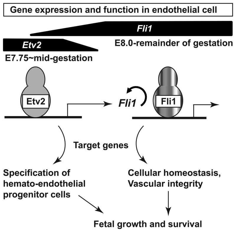
Rationale: Cardiovascular health depends on proper development and integrity of blood vessels. Ets variant 2 (Etv2), a member of the E26 transforming-specific family of transcription factors, is essential to initiate a transcriptional program leading to vascular morphogenesis in early mouse embryos. However, endothelial expression of the Etv2 gene ceases at midgestation; therefore, vascular development past this stage must continue independent of Etv2.

Objective: To identify molecular mechanisms underlying transcriptional regulation of vascular morphogenesis and homeostasis in the absence of Etv2.

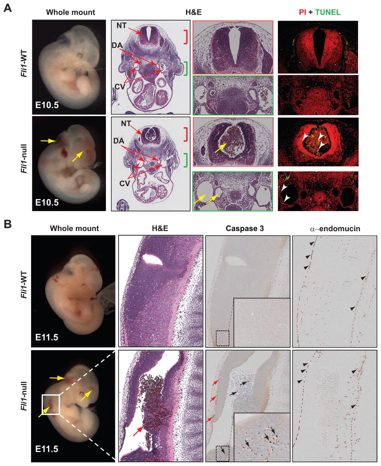
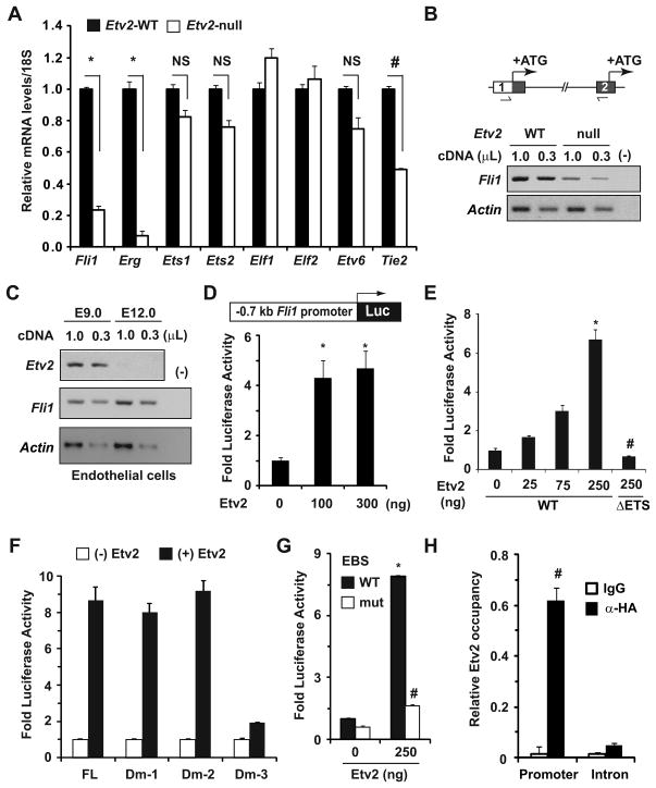
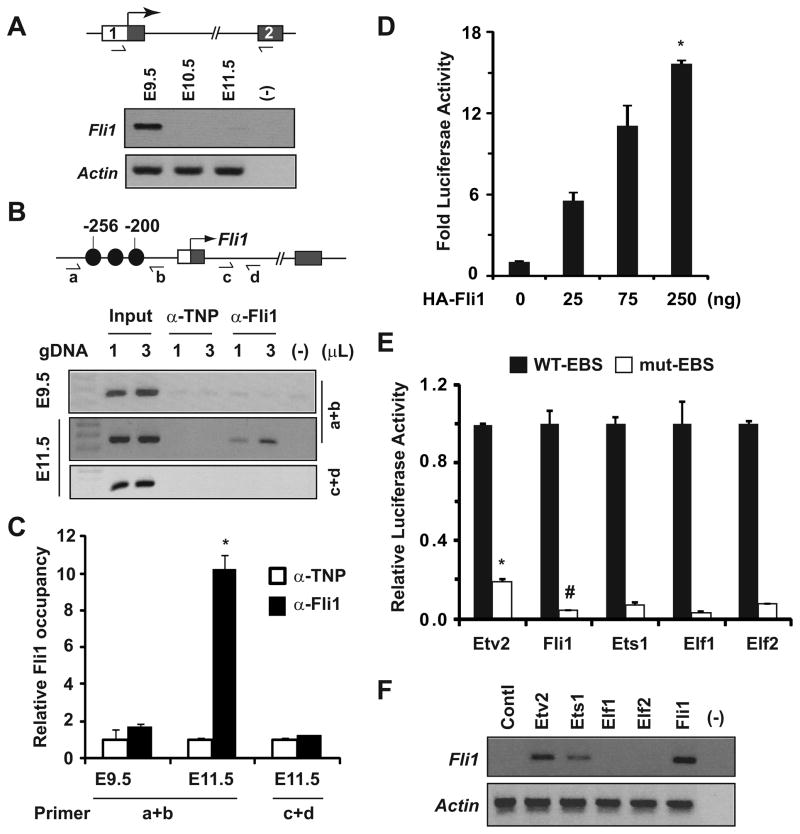
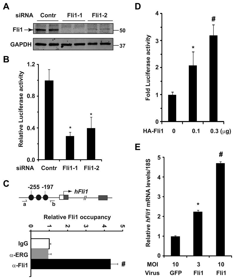
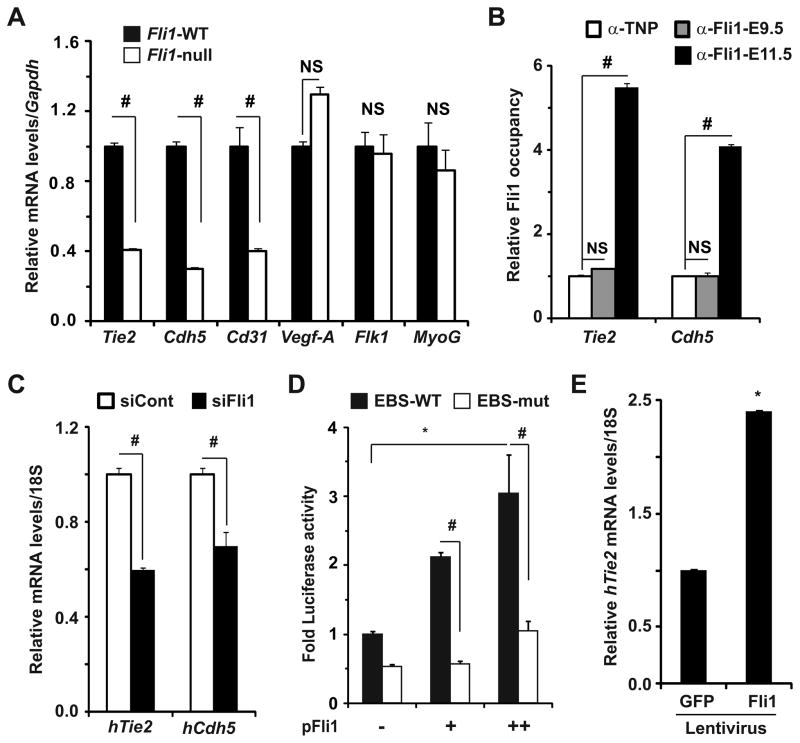
Methods and results: Using loss- and gain-of-function strategies and a series of molecular techniques, we identify Friend leukemia integration 1 (Fli1), another E26 transforming-specific family transcription factor, as a downstream target of Etv2. We demonstrate that Etv2 binds to conserved Ets-binding sites within the promoter region of the Fli1 gene and governs Fli1 expression. Importantly, in the absence of Etv2 at midgestation, binding of Etv2 at Ets-binding sites in the Fli1 promoter is replaced by Fli1 protein itself, sustaining expression of Fli1 as well as selective Etv2-regulated endothelial genes to promote endothelial cell survival and vascular integrity. Consistent with this, we report that Fli1 binds to the conserved Ets-binding sites within promoter and enhancer regions of other Etv2-regulated endothelial genes, including Tie2, to control their expression at and beyond midgestation.

Conclusions: We have identified a novel positive feed-forward regulatory loop in which Etv2 activates expression of genes involved in vasculogenesis, including Fli1. Once the program is activated in early embryos, Fli1 then takes over to sustain the process in the absence of Etv2.

Keywords: apoptosis; developmental biology; embryonic development; gene expression; homeostasis; transcription factors.


Figures

Figure 1
Figure 1. Vascular leakage in Fli1-null embryos at mid-gestation is due to excessive ECs death
(A) Gross (whole-mount) and hematoxylin and eosin (H&E) sections of E10.5 WT and Fli1-null littermates are shown. Blood in the canal of neural tube (NT) but absent in the vasculature, dorsal aorta (DA) and cardinal vein (CV), due to hemorrhage (yellow arrows) is evident in null mice. Histology corresponding to NT (red bracket) and vasculatures (green bracket) are shown to demonstrate increased apoptotic cell death (TUNEL) of ECs, blood and neuropil (arrowheads) in null mice. (B) Gross and histologic (H&E) sections of E11.5 WT and Fli1-null littermates are shown. Higher magnification H&E images of NT illustrate hemorrhage in Fli1-null mice (red arrow). IHC analyses for activated caspase 3 and endomucin revealed increased apoptotic EC death (small red arrows), blood (black arrows) and cells of the neuropil (arrows in inset).
Figure 2
Figure 2. Etv2 governs Fli1 gene expression in vitro and in vivo
(A) qRT-PCR analyses for transcript levels of the indicated genes in E8.0 WT and Etv2-null embryos (n=3–4). Relative gene expression in WT embryos was normalized to 1. Note the significant attenuation of Fli1, Erg (*p<0.001 vs. WT) and Tie2 (#p<0.0001 vs. WT) gene expression in Etv2-null mice. NS: not significant. (B) Semi-quantitative RT-PCR (sqRT-PCR) analyses of Fli1 transcripts spanning exon 1 UTR and exon 2 (boxes) of the Fli1 gene (schematized top) in E8.0 WT and Etv2-null embryos. Translation initiation sites (ATG) in exon 1 and exon 2 are indicated. PCR loading (α-actin) and negative (−) controls are indicated. (C) sqRT-PCR analyses of Etv2 and Fli1 transcripts in GFP+ ECs isolated at the indicated developmental stages. Note the transient co-expression of the Etv2 and Fli1 genes in ECs of early embryos, while expression of the Fli1 gene persisted beyond mid-gestation. (D) Schematic of the Fli1 promoter region (−0.7kb) fused to a luciferase (Luc) reporter. Co-transfection of reporter and indicated amounts of Etv2 expression plasmids in HAECs resulted in significant induction of luciferase activity (*p<0.008 vs. control). (E) Transcriptional assays in COS1 cells elicited dose-dependent activation of luciferase activity by WT, but not mutant (lack of DNA-binding domain, ΔETS) Etv2. (F and G) Fold-activation of luciferase activity in the absence (−) and presence (+) of Etv2 (250ng) from FL and indicated Dm reporter plasmids are shown in panel F. Note the EBSs located between 200–250bp upstream of ATG were essential for efficient activation of luciferase activity, and that activity was significantly abrogated when they were mutated, panel G (*p<0.001 vs. control; # p<0.001 vs. WT). Luciferase activity without Etv2 was normalized to 1. (H) Quantitative analyses of Etv2 occupancy of the Fli1 promoter. Note the specific and significant induction of Etv2 occupancy of the Fli1 promoter (# p<0.001 vs. control).
Figure 3
Figure 3. Fli1 governs Fli1 gene expression via positive autoregulation
(A) Semi-quantitative RT-PCR analyses of Fli1 expression in Fli1-null embryos isolated at the indicated stages. Note the Fli1 transcripts detected at E9.5 were significantly attenuated at mid-gestation. (B and C) ChIP assays (B) and quantitative analyses (C) for Fli1 occupancy of the Fli1 promoter (schematized top) in embryos isolated at the indicated stages. Fli1-DNA complexes were immunoprecipitated with anti-Fli1 and anti-TNP (control) sera. Note that promoter-specific Fli1 binding was enriched at E11.5, but not at E9.5, embryos (*p<0.0001 vs. control). (D) Transcriptional assays using Dm-2 reporter and Fli1 expression plasmids in COS1 cells elicited robust and dose-dependent activation of luciferase activity (*p<0.0001 vs. control). (E) Transcriptional assays in the presence of indicated ETS factors (250ng) and Dm-2 reporter plasmids harboring WT and mutated EBSs are shown. Note that mutation of EBSs significantly attenuated luciferase activity. Luciferase activity for each ETS factor from reporter plasmid harboring WT EBS was normalized to 1. (F) RT-PCR analyses of the endogenous Fli1 transcripts following expression of the indicated ETS factors in C2C12 cells. Note that Etv2 and Fli1, but not Elf1 or Efl2, induced endogenous Fli1 transcript levels.
Figure 4
Figure 4. Fli1 is required for sustained Fli1 expression for the remainder of gestation
(A) Western blot analyses of Fli1 in HAECs transfected with control (Contr) and Fli1-specific siRNAs. Note the marked reduction of Fli1 protein levels in siFli1-treated cells. GAPDH was used as a loading control. Numbers indicates protein molecular markers (kD). (B) Baseline Fli1 reporter activity in HAECs transfected with the indicated siRNA (p<0.05 vs. control). (C) ChIP-qPCR analyses of Fli1 occupancy of the Fli1 promoter. Note the Fli1, but not Erg, occupancy of Fli1 promoter (# p<0.003 vs. control). (D) Transcriptional assays reveal significant and dose-dependent induction of luciferase activity by Fli1 from Fli1 reporter plasmid in HAECs (*p<0.001 vs. control). (E) qRT-PCR analyses for the endogenous Fli1 transcripts in HAECs infected with control (GFP) and Fli1-encoding lentivirus (*p<0.003 vs. control, #p<0.0001 vs. control).
Figure 5
Figure 5. Fli1 governs selective Etv2-regulated endothelial gene expression at and beyond mid-gestation
(A) qRT-PCR analyses for transcript levels of the indicated genes in E10.5 WT and Fli1-null embryos (n=3). Relative gene expression in WT mice was normalized to 1 (*p<0.001 vs. WT). NS: not significant. (B) ChIP-qPCR analyses demonstrating Fli1 occupancy of the promoter of indicated endothelial genes in E11.5, but not E9.5, embryos (# p<0.001 vs. control). (C) qRT-PCR analyses revealing significant attenuation of Tie2 and Cdh5 transcript levels in siFli1-treated HAECs (#p<0.01 vs. control). (D) Transcriptional assays in COS1 cells reveal significant and dose-dependent induction of luciferase activity by Fli1 from Tie2-UPF reporter plasmid harboring WT (black), but not mutated (white), EBSs (*p<0.001 vs. control; # p<0.002 vs. WT EBS). (E) qRT-PCR analyses for Tie2 transcript levels in HAECs infected with control (GFP) and Fli1-encoding lentivirus (MOI 10) (*p<0.001 vs. control).
Figure 6
Figure 6
Working model for positive autoregulation between Etv2 and Fli1 regulates vascular morphogenesis and integrity at early and mid-gestation of mouse embryogenesis. Embryonic expression of Etv2 and Fli1 genes at distinct developmental stages and their role in EC are indicated. See text for details.

References

    1. Flammer AJ, Anderson T, Celermajer DS, Creager MA, Deanfield J, Ganz P, Hamburg NM, Luscher TF, Shechter M, Taddei S, Vita JA, Lerman A. The assessment of endothelial function: From research into clinical practice. Circulation. 2012;126:753–767. - PMC - PubMed
    1. Olson EN, Srivastava D. Molecular pathways controlling heart development. Science. 1996;272:671–676. - PubMed
    1. Choi K. The hemangioblast: A common progenitor of hematopoietic and endothelial cells. J Hematother Stem Cell Res. 2002;11:91–101. - PubMed
    1. Chung YS, Zhang WJ, Arentson E, Kingsley PD, Palis J, Choi K. Lineage analysis of the hemangioblast as defined by Flk1 and Scl expression. Development. 2002;129:5511–5520. - PubMed
    1. De Val S, Black BL. Transcriptional control of endothelial cell development. Dev Cell. 2009;16:180–195. - PMC - PubMed

Publication types

MeSH terms

LinkOut - more resources