Review
. 2014 Oct;1839(10):1030-1045.
doi: 10.1016/j.bbagrm.2014.04.005. Epub 2014 Apr 13.

Single-molecule studies of riboswitch folding

Affiliations
Review

Single-molecule studies of riboswitch folding

Andrew Savinov et al. Biochim Biophys Acta. 2014 Oct.

Abstract

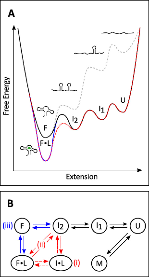
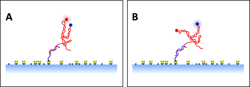
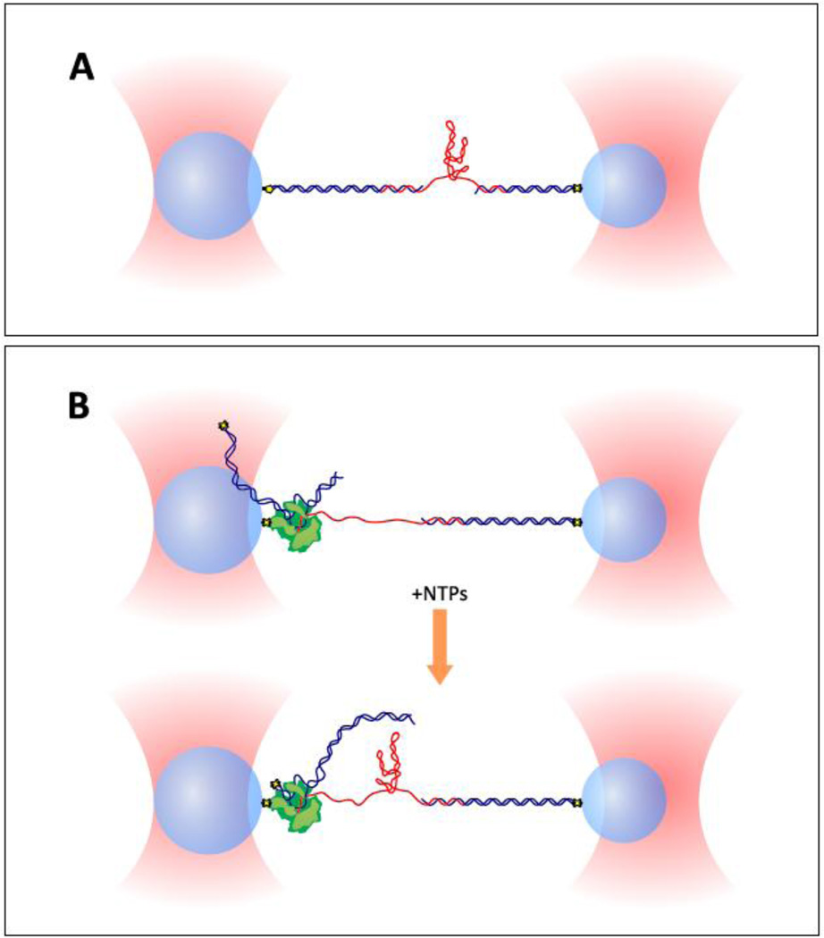
The folding dynamics of riboswitches are central to their ability to modulate gene expression in response to environmental cues. In most cases, a structural competition between the formation of a ligand-binding aptamer and an expression platform (or some other competing off-state) determines the regulatory outcome. Here, we review single-molecule studies of riboswitch folding and function, predominantly carried out using single-molecule FRET or optical trapping approaches. Recent results have supplied new insights into riboswitch folding energy landscapes, the mechanisms of ligand binding, the roles played by divalent ions, the applicability of hierarchical folding models, and kinetic vs. thermodynamic control schemes. We anticipate that future work, based on improved data sets and potentially combining multiple experimental techniques, will enable the development of more complete models for complex RNA folding processes. This article is part of a Special Issue entitled: Riboswitches.

Keywords: Gene regulation; Optical trap; Optical tweezers; Regulatory mechanism; Single molecule.


Figures

Figure 1
Figure 1. Riboswitch aptamer folding
(A) Notional free energy landscape for the folding and ligand binding of a generic riboswitch aptamerdomain. Energy is plotted vs. end-to-end extension, the reaction coordinate used in optical trappingexperiments. Example conformational states (U: unfolded, I1,2: intermediates; F: folded, F•L: folded andbound to ligand) are shown for each energy well (bound ligand in green). Ligand-free energy landscapesat zero applied load (gray dashed curve) and under external tension (black curve) are plotted againstextension. Also shown are folding landscapes in the presence of ligand, following either a conformational selection (blue curve) or an induced fit (red dotted curve) model for binding, both under applied tension. (B) Kinetic scheme matching the energy landscape in (A), with additional potential states represented(M: a misfolded state, I•L: an induced-fit intermediate). Roman numerals indicate two possible induced-fit pathways (i, ii) and the conformational selection pathway (iii).
Figure 2
Figure 2. Typical experimental geometry for a surface-based smFRET assay of riboswitch folding
The riboswitch (red) is anchored to a treated glass surface (light blue shading) via a single-stranded DNA handle (blue), which is specifically attached to the treated surface via chemical linkers (black dots, yellow crosses). Conformational changes alter the proximity of the donor and acceptor fluorophores (blue and red stars), producing a change in the FRET efficiency. Panel (A) shows close proximity of the donor and acceptor, which results in a high FRET state. Panel (B) shows a molecular conformation in which the donor and acceptor are separated, resulting in a low FRET state.
Figure 3
Figure 3. Two dual-beam optical trap assays for riboswitch folding
Dual optical traps (salmon) hold functionalized polystyrene beads (light blue spheres) which are each attached to a DNA handle (blue) or RNA polymerase (green) via specific attachment chemistries (yellow and black shapes). The riboswitch RNA (red) is hybridized to single-stranded overhang regions at the ends of double-stranded DNA handles. The end-to-end extension of the riboswitch construct can be measured and controlled with nanometer-level precision. Panel (A) shows a geometry for studying riboswitch constructs synthesized in vitro. Panel (B) describes a setup for studying riboswitches transcribed in situ by RNA polymerase from a DNA template capped with a “roadblock” (yellow and black shapes); addition of NTPs triggers elongation of the nascent RNA (upper image), producing the riboswitch construct (lower image). The geometry in (B) allows the study of co-transcriptional as well as post-transcriptional RNA folding.
Figure 4
Figure 4. Riboswitches studied by single-molecule techniques
Secondary structures are represented, with ligand (green oblong) binding represented schematically and key tertiary interactions in the ligand-bound folded state indicated notionally in orange or as pseudoknot basepairs (G, H). (A) pbuE adenine riboswitch aptamer; (B) add adenine riboswitch aptamer; (C) xpt guanine riboswitch aptamer; (D) thiC TPP riboswitch aptamer; (E) thiM TPP riboswitch aptamer;(F) metI SAM class I riboswitch aptamer; (G) metX SAM class II riboswitch; (H) left: preQ1 class IIriboswitch, right: preQ1 class I riboswitch; (I) tfoX c-di-GMP class I riboswitch aptamer; (J) lysC lysineriboswitch aptamer. FRET labeling sites are indicated with blue and salmon dots (triangles, squares andstars indicate additional possible labeling sites) for the donor and acceptor locations, respectively.

References

    1. Anfinsen CB. Principles that govern folding of protein chains. Science. 1973;181:223–230. - PubMed
    1. Anfinsen CB, Haber E, Sela M, White FH. Kinetics of formation of native ribonuclease during oxidation of reduced polypeptide chain. Proc Natl Acad Sci U S A. 1961;47:1309–1314. - PMC - PubMed
    1. Rother K, Rother M, Boniecki M, Puton T, Bujnicki JM. RNA and protein 3D structure modeling: similarities and differences. J Mol Model. 2011;17:2325–2336. - PMC - PubMed
    1. Laing C, Schlick T. Computational approaches to RNA structure prediction, analysis, and design. Curr Opin Struct Biol. 2011;21:306–318. - PMC - PubMed
    1. Zhang Y. Progress and challenges in protein structure prediction. Curr Opin Struct Biol. 2008;18:342–348. - PMC - PubMed

LinkOut - more resources