. 2014 Jun;44(7):485-96.
doi: 10.1016/j.ijpara.2014.03.005. Epub 2014 Apr 12.

Hc-daf-2 encodes an insulin-like receptor kinase in the barber's pole worm, Haemonchus contortus, and restores partial dauer regulation

Affiliations

Hc-daf-2 encodes an insulin-like receptor kinase in the barber's pole worm, Haemonchus contortus, and restores partial dauer regulation

Facai Li et al. Int J Parasitol. 2014 Jun.

Abstract

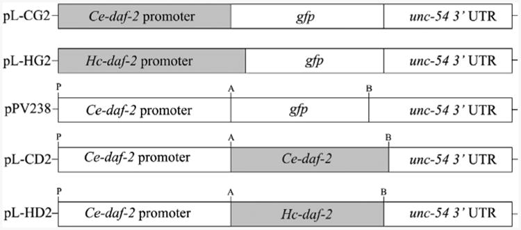
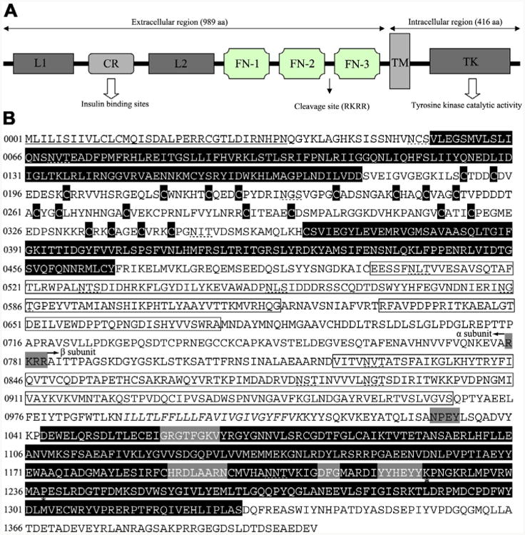
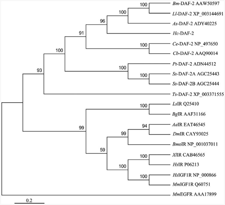
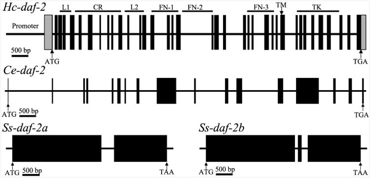
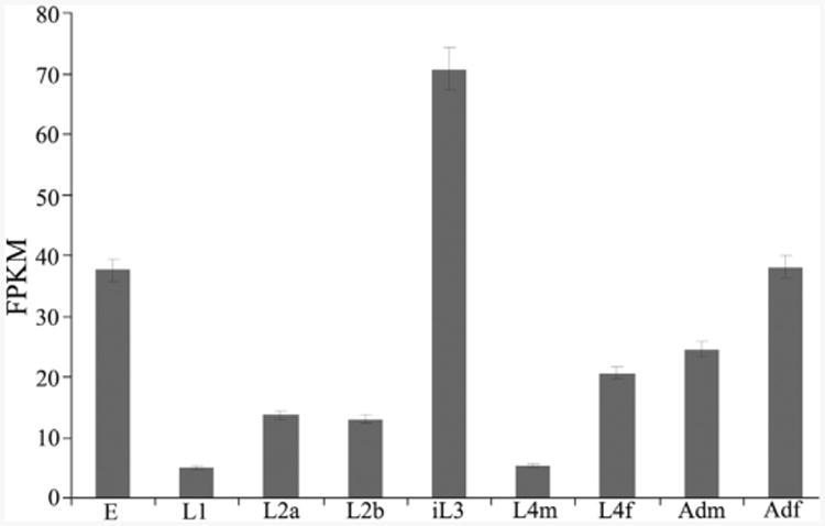
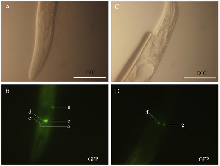
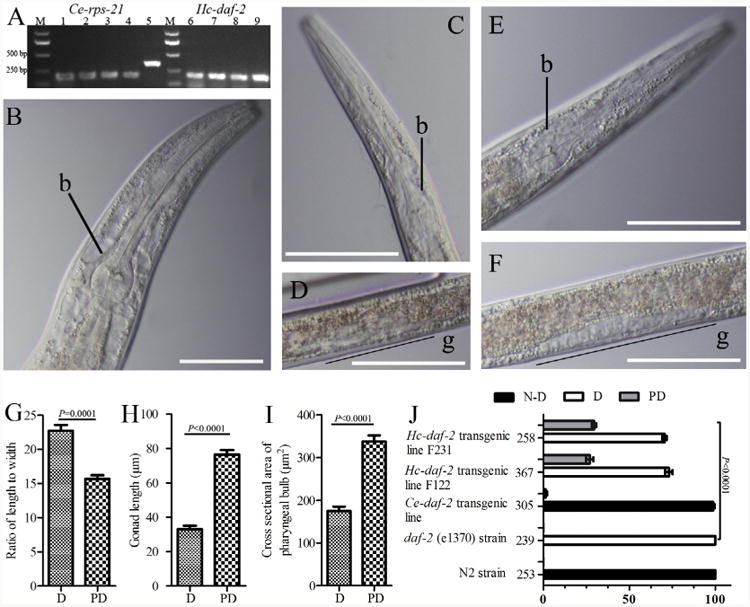
Infective L3s (iL3s) of parasitic nematodes share common behavioural, morphological and developmental characteristics with the developmentally arrested (dauer) larvae of the free-living nematode Caenorhabditis elegans. It is proposed that similar molecular mechanisms regulate entry into or exit from the dauer stage in C. elegans, and the transition from free-living to parasitic forms of parasitic nematodes. In C. elegans, one of the key factors regulating the dauer transition is the insulin-like receptor (designated Ce-DAF-2) encoded by the gene Ce-daf-2. However, nothing is known about DAF-2 homologues in most parasitic nematodes. Here, using a PCR-based approach, we identified and characterised a gene (Hc-daf-2) and its inferred product (Hc-DAF-2) in Haemonchus contortus (a socioeconomically important parasitic nematode of ruminants). The sequence of Hc-DAF-2 displays significant sequence homology to insulin receptors (IR) in both vertebrates and invertebrates, and contains conserved structural domains. A sequence encoding an important proteolytic motif (RKRR) identified in the predicted peptide sequence of Hc-DAF-2 is consistent with that of the human IR, suggesting that it is involved in the formation of the IR complex. The Hc-daf-2 gene was transcribed in all life stages of H. contortus, with a significant up-regulation in the iL3 compared with other stages. To compare patterns of expression between Hc-daf-2 and Ce-daf-2, reporter constructs fusing the Ce-daf-2 or Hc-daf-2 promoter to sequence encoding GFP were microinjected into the N2 strain of C. elegans, and transgenic lines were established and examined. Both genes showed similar patterns of expression in amphidial (head) neurons, which relate to sensation and signal transduction. Further study by heterologous genetic complementation in a daf-2-deficient strain of C. elegans (CB1370) showed partial rescue of function by Hc-daf-2. Taken together, these findings provide a first insight into the roles of Hc-daf-2/Hc-DAF-2 in the biology and development of H. contortus, particularly in the transition to parasitism.

Keywords: Development; Haemonchus contortus; Parasitic nematode; Transgenesis; daf-2.


Figures

Fig. 1
Fig. 1
Cloning strategy for reporter and rescuing constructs. The constructs containing the Caenorhabditis elegans Ce-daf-2 promoter (pL-CG2) and the Haemonchus contortus Hc-daf-2 promoter (pL-HG2) were made based on pPD95.75 by overlap extension PCR. Rescuing constructs containing coding sequences of Ce-daf-2 (pL-CD2) and Hc-daf-2 (pL-HD2) were made by removing the gfp coding sequence from pPV238 (Massey et al., 2013) and linking the appropriate cDNA. A, B and P represent the restriction sites for AgeI, BstZ17I and PstI, respectively. UTR, untranslated region.
Fig. 2
Fig. 2
Insulin-like receptor of Haemonchus contortus consisting of representative structural domains. (A) Domain structure of the insulin-like receptor of Haemonchus contortus. Analysis of the amino acid sequence for the insulin-like receptor predicted all of the characteristic domains of insulin-like receptors from other taxa, including ligand binding loop 1 (L1) and loop 2 (L2); the cysteine-rich region (CR); three fibronectin type domains (FN-1, FN-2 and FN-3); the transmembrane domain (TM); and a tyrosine kinase domain (TK) (Konrad et al., 2003). (B) Protein sequence and structural features of the insulin-like receptor of H. contortus (Hc-DAF-2). The putative receptor L domains, cysteine residues of the CR domain and a TK domain are black boxes with white lettering, the FN domains are boxed. The TM region is italicised and conserved domains (GXGXXG, HRDLAARN, DFG and YXXXYY) (White et al., 1988) are boxed by a grey background. Further indicated are the potential proteolytic cleavage site RKRR (grey box) which divided the Hc-DAF-2 into a subunit and β subunit as well as the juxtamembrane NPxY and the Mg2+ binding domain. Two residues (K1224 and P1238) are marked by asterisks, which are highly conserved in the activation loop and might be involved in interaction with downstream signalling proteins (Hubbard, 1997; Massey et al., 2013). The signal peptide is underlined and the putative N-linked glycosylation sites (NxT/S) are marked with dashed lines.
Fig. 3
Fig. 3
A rooted neighbour joining tree showing the relationships of Haemonchus contortus insulin-like receptor Hc-DAF-2 to the insulin-like receptors of nine nematodes and nine non-nematodes. The tree was calculated using the Jones-Taylor-Thornton model in the MEGA program version 5.0. Bootstrap values above or below the branches (1000 iterations) are shown for robust clades (>50%). These 18 species include nine nematodes (Brugia malayi, Bm-DAF-2; Loa loa, Ll-DAF-2; Ascaris suum, As-DAF-2; Caenorhabditis elegans, Ce-DAF-2; Caenorhabditis briggsae, Cb-DAF-2; Parastrongyloides trichosuri, Pt-DAF-2; Strongyloides stercoralis, Ss-DAF-2A and Ss-DAF-2B; Trichinella spiralis, Ts-DAF-2), Molluscs (Lymnaea stagnalis, LsIR; Biomphalaria glabrata, BgIR), Insects (Aedes aegypti, AaIR; Drosophila melanogaster, DmIR; Bombyx mori, BmoIR), Vertebrates (Xenopus laevis, XlIR; Homo sapiens, HsIR; Homo sapiens, HsIGF1R; Mus musculus, MmIGF1R). Their corresponding accession numbers are listed on the right of each species. Mus musculus EGFR (MmEGFR) was used as the outgroup.
Fig. 4
Fig. 4
Schematic diagram showing the genomic organization of daf-2 from Haemonchus contortus (Hc-daf-2), Caenorhabditis elegans (Ce-daf-2) (Kimura et al., 1997) and Strongyloides stercoralis (Ss-daf-2a and Ss-daf-2b) (Massey et al., 2013). Black boxes represent exons. The lines between the exons represent introns. The structural domains encoded by Hc-daf-2 are marked on the top of diagram of Hc-daf-2 gene, located to the corresponding positions. Arrows indicate the start (ATG) or stop codon (TGA). L, ligand binding loop; CR, cysteine-rich region; FN, fibronectin type domains; TM, transmembrane domain; TK, tyrosine kinase domain.
Fig. 5
Fig. 5
Transcriptional profile of Hc-daf-2 in different developmental stages of Haemonchus contortus. Transcript abundances were compared in eight developmental stages, each in biological triplicate (n = 3). Eight developmental stages as follows: eggs (E), L1, L2, infective L3 (iL3), the L4 males (L4m), the L4 females (L4f); adult males (Adm), adult females (Adf). Transcript abundances were counted as fragments per kilobase of coding exon per million mapped reads (FPKM). Error bars represent 95% confidence intervals.
Fig. 6
Fig. 6
Representative expression profiles displayed in Caenorhabditis elegans using two GFP constructs, pL-CG2 and pL-HG2 (cf. Fig. 1). (A and B) Differential interference contrast (DIC) and fluorescence images of a N2 (wild type) L3 using the construct Ce-daf-2 p∷gfp (pL-CG2), respectively. GFP reporter expression was present in the head neuron (a), amphidial neurons including ASH (b), ADF (d) and AWA (e), and nerve ring (c). (C and D) DIC and fluorescence images showing the expression of construct Haemonchus contortus Hc-daf-2 p∷gfp (pL-HG2) in L3 stage of a N2 C. elegans. GFP reporter expression was present in amphidial neuron AWA (f and g). Scale bars = 50 μm.
Fig. 7
Fig. 7
Results of dauer developmental assays on mutant and transgenic Caenorhaboditis elegans (Ce) strains. (A) Reverse transcriptase (RT)-PCR to detect the transcription of Haemonchus contortus Hc-daf-2-specific mRNA in daf-2 (e1370) transgenic lines. The constitutively expressed mRNA encoding the ribosomal protein small subunit RPS-21 (Ce-rps-21) was used as a loading control. The Ce-daf-2 transgenic lines were transformed with pL-CD2, and the Hc-daf-2 transgenic lines were transformed with pL-HD2. RNA was extracted from the transgenic worms exhibiting the roller phenotype. Templates are as follows: lanes 1–4 or 6–9 represent Hc-daf-2 transformed lines F121, F122, F231, F232, respectively; lane 5 is C. elegans genomic DNA; M, bp size marker. (B - J) Results of dauer-switching assays on well-fed worms from genetic line N2 and daf-2 (e1370) mutants transformed with the indicated transgenes as differential interference contrast (DIC) images. b, bulb; g, gonad. Scale bars (B–F) = 50 lm. (B) Caenorhaboditis elegans transgenic line expressing Ce-daf-2 mRNA in a daf-2 (e1370) mutant strain; (C and D) C. elegans dauer from a daf-2 (e1370) strain; (E and F) C. elegans transgenic line expressing Hc-daf-2 mRNA in a daf-2 (e1370) mutant strain; (G) ratio of body length to body width between dauer (D) and partial dauer (PD) larvae; (H) comarpison of gonad length between dauer and partial dauer larvae; (I) cross-sectional area of the pharyngeal bulb between dauer and partial dauer larvae; (J) percentage of total population in transgenic lines and controls. Numbers on the left of the X-axis represent the total number of worms counted for each line. P ≤ 0.0001 denotes a statistically significant difference between dauer, partial dauer and non-dauer (N-D) larval development in the parental daf-2 (e1370) strain.

References

    1. Aboobaker AA, Blaxter ML. Functional genomics for parasitic nematodes and platyhelminths. Trends Parasitol. 2004;20:178–184. - PubMed
    1. Apfeld J, Kenyon C. Cell nonautonomy of C. elegans daf-2 function in the regulation of diapause and life span. Cell. 1998;95:199–210. - PubMed
    1. Ayyadevara S, Tazearslan C, Bharill P, Alla R, Siegel E, Shmookler Reis RJ. Caenorhabditis elegans PI3K mutants reveal novel genes underlying exceptional stress resistance and lifespan. Aging Cell. 2009;8:706–725. - PMC - PubMed
    1. Bargmann CI. Chemosensation in C. elegans. WormBook, editor. The C elegans Research Community. 2006 Available from: < http://www.wormbook.org/chapters/www_chemosensation/chemosensation.html>. - PMC - PubMed
    1. Bargmann CI, Horvitz HR. Chemosensory neurons with overlapping functions direct chemotaxis to multiple chemicals in C. elegans. Neuron. 1991;7:729–742. - PubMed

Publication types