. 2014 Apr 10;19(4):4433-51.
doi: 10.3390/molecules19044433.

The effect of chitin size, shape, source and purification method on immune recognition

Affiliations

The effect of chitin size, shape, source and purification method on immune recognition

Francisco J Alvarez. Molecules. .

Abstract

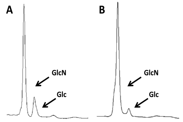
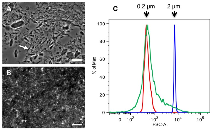
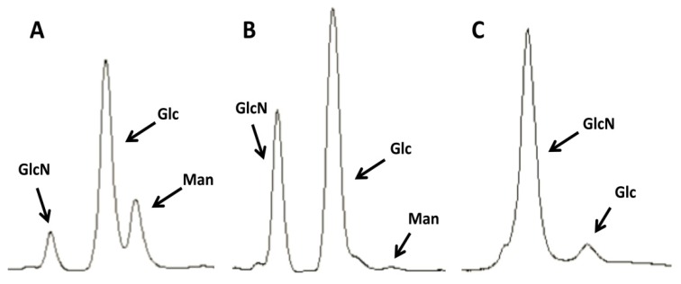
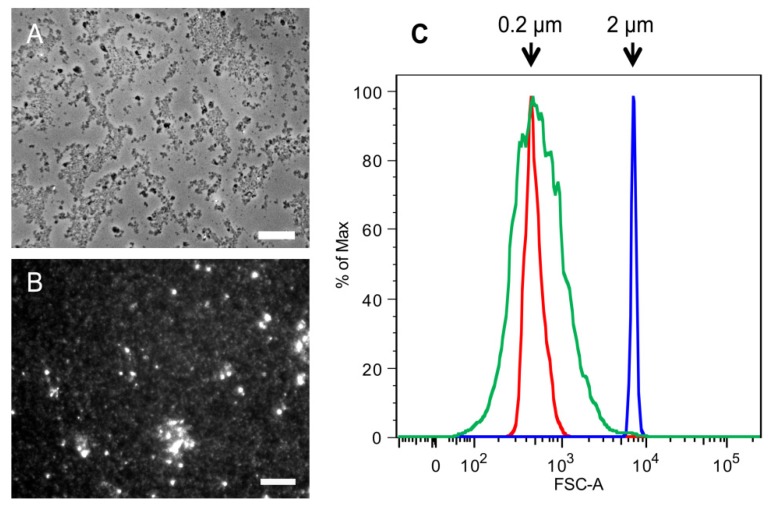
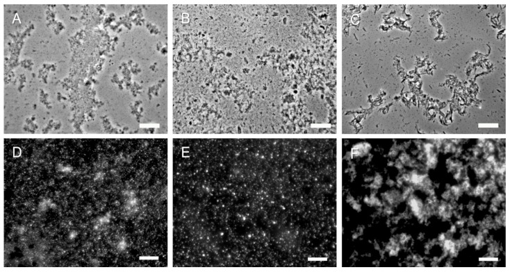
The animal immune response to chitin is not well understood and needs to be investigated further. However, this is a challenging topic to study because of the technical difficulties in purifying chitin, and because this material usually comes associated with contaminating components that can activate the immune system. In this study, improvements to previously described purification protocols were investigated for chitin obtained from different sources, including commercial shellfish, Candida albicans yeast and hyphal cell walls, as well as cell walls of the filamentous fungi Aspergillus fumigatus and Mucor circinelloides. The immune response to these different chitin preparations was tested using human peripheral blood mononuclear cells. In agreement with previous literature, small chitin particles of an average size of 0.2 µm were not immunogenic. On the other hand, bigger chitin particles induced in some cases a pro-inflammatory response. The results of this work suggest that not only the purity and size of the chitin particles, but also their shape can influence immune recognition.


Conflict of interest statement

The author declares no conflicts of interest.

Figures

Figure 1
Figure 1
Purity of commercial crab chitin before and after treatment under alkali and acid conditions. Analysis of (A) untreated commercial crab chitin or (B) the same chitin that was treated by boiling twice with 5% KOH and then three times in a mix of acetic acid and H2O2. Samples were then hydrolyzed with TFAA and then the carbohydrate content was analyzed by HPLC. GlcN indicates glucosamine that results from the deacetylation of the GlcNAc under the hydrolysis conditions. Glc indicates the presence of glucose from contaminating glucans, which were not completely eliminated by the chemical treatments.
Figure 2
Figure 2
Comparison of SSCs and 2 k particles obtained from commercial crab chitin. Microscopic analysis of (A) 2 k chitin viewed by phase contrast microscopy; and (B) super small chitin particles (SSCs) that were stained with CFW and viewed by fluorescence microscopy; (C) Flow Cytometry analysis of SSCs. Red and blue indicate the profiles of 0.2 and 2 µm standard beads, respectively; green indicates the profile of SSCs. Arrow in (A) points to a spike-shaped chitin particle. Bars, 10 µm.
Figure 3
Figure 3
Purification of chitin from C. albicans yeast phase cells. Images representative of the HPLC profiles of C. albicans extracts at different stages of the chitin purification process. (A) Untreated cell walls; (B) after 3 KOH boils; (C) after an additional boil in a 1:1 mix of acetic acid and 40% H2O2. Man, mannose (mannans); Glc, glucose (glucans); GlcN, glucosamine (chitin).
Figure 4
Figure 4
Comparison of SSCs and 2 k chitin particles from C. albicans yeast cells.Microscopic analysis of (A) 2 k chitin viewed by phase contrast microscopy; and (B) SSCs viewed by fluorescence microscopy after CFW staining; (C) Flow Cytometry analysis of SSCs. Red and blue indicate the profiles of 0.2 and 2 µm standard beads, respectively; green indicate the profile of SSCs. Bars, 10 µm.
Figure 5
Figure 5
Representative microscopy images of SSCs and 2 k chitin from hyphae of three different fungi. (AC) 2 k fractions from C. albicans, A. fumigatus and M. circinelloides, respectively (phase contrast); (DF) SSCs from the corresponding fungi were stained with CFW and then viewed by fluorescence microscopy. Bars, 10 µm.

References

    1. Blumenthal H.J., Roseman S. Quantitative estimation of chitin in fungi. J. Bacteriol. 1957;74:222–224. - PMC - PubMed
    1. Wagner G.P., Lo J., Laine R., Almeder M. Chitin in the epidermal cuticle of a vertebrate (paralipophrys trigloides, blenniidae, teleostei) Experientia. 1993;49:317–319. doi: 10.1007/BF01923410. - DOI
    1. Kurita K. Chitin and chitosan: Functional biopolymers from marine crustaceans. Mar. Biotechnol. 2006;8:203–226. doi: 10.1007/s10126-005-0097-5. - DOI - PubMed
    1. Jang M.-K., Kong B.-G., Jeong Y.-I., Lee C.H., Nah J.-W. Physicochemical characterization of alpha-chitin, beta-chitin, and gamma-chitin separated from natural resources. J. Polym. Sci. 2004;42:3423–3432. doi: 10.1002/pola.20176. - DOI
    1. Khoushab F., Yamabhai M. Chitin research revisited. Mar. Drugs. 2010;8:1988–2012. doi: 10.3390/md8071988. - DOI - PMC - PubMed

Publication types

MeSH terms