. 2014 May 30;289(22):15739-49.
doi: 10.1074/jbc.M113.524009. Epub 2014 Apr 10.

Solution NMR structure of the DNA-binding domain from Scml2 (sex comb on midleg-like 2)

Affiliations

Solution NMR structure of the DNA-binding domain from Scml2 (sex comb on midleg-like 2)

Irina Bezsonova. J Biol Chem. .

Abstract

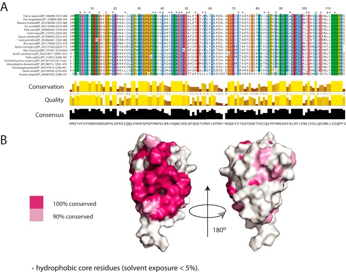
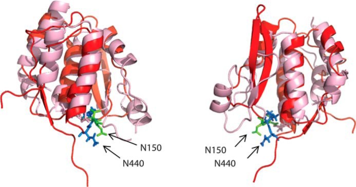
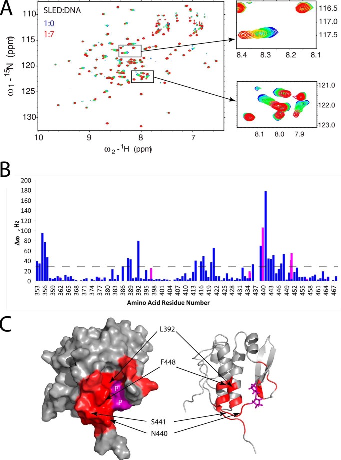
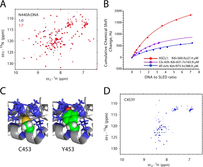
Scml2 is a member of the Polycomb group of proteins involved in epigenetic gene silencing. Human Scml2 is a part of a multisubunit protein complex, PRC1 (Polycomb repressive complex 1), which is responsible for maintenance of gene repression, prevention of chromatin remodeling, preservation of the "stemness" of the cell, and cell differentiation. Although the majority of PRC1 subunits have been recently characterized, the structure of Scml2 and its role in PRC1-mediated gene silencing remain unknown. In this work a conserved protein domain within human Scml2 has been identified, and its structure was determined by solution NMR spectroscopy. This module was named Scm-like embedded domain, or SLED. Evolutionarily, the SLED domain emerges in the first multicellular organisms, consistent with the role of Scml2 in cell differentiation. Furthermore, it is exclusively found within the Scm-like family of proteins, often accompanied by malignant brain tumor domain (MBT) and sterile α motif (SAM) domains. The domain adopts a novel α/β fold with no structural analogues found in the Protein Data Bank (PDB). The ability of the SLED to bind double-stranded DNA was also examined, and the isolated domain was shown to interact with DNA in a sequence-specific manner. Because PRC1 complexes localize to the promoters of a specific subset of developmental genes in vivo, the SLED domain of Scml2 may provide an important link connecting the PRC1 complexes to their target genes.

Keywords: DNA-binding Protein; Epigenetics; NMR; Polycomb; Protein Structure.


Figures

FIGURE 1.
FIGURE 1.
SLED domain in human proteins. A, schematic representation of SLED-containing proteins in humans. SLED, MBT, and SAM domains are shown in red, cyan, and black, respectively. B, multiple sequence alignment of SLED domains from human proteins performed using ClustalW (29). Invariant residues are shown in red. The predicted secondary structure elements of the domain are shown above the Scml2 sequence. Scml2 cysteine residues are marked with an asterisk.
FIGURE 2.
FIGURE 2.
SLED domain conservation in multicellular organisms. A phylogenetic tree of SLED domain containing proteins is shown. Each species is represented by an individual color (right). The domain architecture for each homolog is shown following the name of the protein. SLED, MBT, and SAM domains are shown in red and cyan squares and black circles, respectively. The tree was generated using the Interactive Tree of Life server iTOL (30).
FIGURE 3.
FIGURE 3.
Solution NMR structure of the SLED domain from human Scml2 (residues 354–467). A, NMR ensemble of the 20 lowest energy structures of the human Scml2 SLED domain (backbone representation only). The protein chain is rainbow-colored from blue to red; N and C termini are labeled. B, schematic representation of the SLED domain topology. C, ribbon representation of the SLED domain shown in two orientations; secondary structure elements are labeled. D, charge distribution on the surface of the SLED domain (shown in the same orientation as C).
FIGURE 4.
FIGURE 4.
Amino acid residue conservation in SLED domains. A, multiple sequence alignment of the Scml2 SLED domain across the species. Default ClustalW (29) colors are used for each residue. Residues with conservation of 90% or higher are highlighted, and residues that form the hydrophobic core of the domain are annotated with an asterisk. B, SLED surface conservation. Conserved residues (pink) are mapped on the surface of the human Scml2 SLED. Two orientations are shown to illustrate that conserved residues form a continuous patch on the SLED domain surface. The molecule on the right is rotated by 180° relative to the molecule on the left.
FIGURE 5.
FIGURE 5.
Structural alignment of the Scml2 SLED and SeqA. The SLED domain is shown as a red ribbon, and the SeqA (PDB ID: 1LRR) is shown in pink. The side chains of the SeqA loop residues involved in DNA binding are shown as green sticks (Asn-150, Thr-151), and the side chains of the SLED residues displaying the largest chemical shift changes upon DNA binding are shown as blue sticks (Asn-440, Ser-441).
FIGURE 6.
FIGURE 6.
Scml2 SLED domain and dsDNA binding. A, overlay of 15N/1H HSQC spectra of the free (blue) and DNA-bound SLED domain. The two insets show gradual changes in peak positions (from blue to red) in selected spectral regions upon the addition of ASCL1 dsDNA. B, NMR frequency difference Δω = (ΔωN2 + ΔωH2)½ (at 800 MHz for 1H) between the first and the last points of DNA titration for each amino acid residue of SLED are shown as a histogram. Δω values for backbone HN groups are shown in blue, and those for the HN groups of Gln and Asn side chains are shown in magenta. C, SLED amino acid residues that form the DNA-binding site (with Δω values > 30 Hz) are shown in red on a surface representation (left) and ribbon representation (right) of the SLED structure. Selected DNA-binding residues are labeled. Proline residues within the DNA-binding site are shown in purple on the SLED surface and labeled as P. Titration data for proline residues are not available because they are not present in the 15N/1H HSQC spectrum due to a missing HN group in their chemical structure.
FIGURE 7.
FIGURE 7.
Mutational analysis of Scml2 SLED. A, overlay of 15N/1H HSQC spectra of the free (blue) N440A SLED domain and N440A in the presence of 7 m excess of ASCL1 oligonucleotide (red). B, DNA titration curves derived from the NMR chemical shift perturbation experiments. Cumulative chemical shift changes for all amino acid residues in the domain are plotted as a function of DNA to SLED ratio. Experimental data and fitting curves for three different double-stranded oligonucleotides (dsAGGAGCGGGAG, dsGGGCGCGCCC, and dsTTTATATAAA) are shown in red, purple, and blue, respectively. The resulting Kd values are shown at the bottom of the graph. C, the model of the C453Y mutant of the SLED domain. The packing of the Cys-453 (top) versus Tyr-453 (bottom) side chains within the hydrophobic core is shown. A portion of the protein is shown as a ribbon, and the side chains of the hydrophobic amino acid residues surrounding Cys-453 are shown as sticks. The side-chain atoms of the Cys-453 and Tyr-453 are shown as spheres. D, 15N/1H HSQC spectrum of the C453Y of the Scml2 SLED domain reveals that it is unfolded in solution.

References

    1. Aloia L., Di Stefano B., Di Croce L. (2013) Polycomb complexes in stem cells and embryonic development. Development 140, 2525–2534 - PubMed
    1. Gao Z., Zhang J., Bonasio R., Strino F., Sawai A., Parisi F., Kluger Y., Reinberg D. (2012) PCGF homologs, CBX proteins, and RYBP define functionally distinct PRC1 family complexes. Mol. Cell 45, 344–356 - PMC - PubMed
    1. Simon J. A., Kingston R. E. (2013) Occupying chromatin: Polycomb mechanisms for getting to genomic targets, stopping transcriptional traffic, and staying put. Mol. Cell 49, 808–824 - PMC - PubMed
    1. Wang L., Jahren N., Miller E. L., Ketel C. S., Mallin D. R., Simon J. A. (2010) Comparative analysis of chromatin binding by Sex Comb on Midleg (SCM) and other Polycomb group repressors at a Drosophila Hox gene. Mol. Cell. Biol. 30, 2584–2593 - PMC - PubMed
    1. Bornemann D., Miller E., Simon J. (1998) Expression and properties of wild-type and mutant forms of the Drosophila sex comb on midleg (SCM) repressor protein. Genetics 150, 675–686 - PMC - PubMed

Publication types

MeSH terms

Associated data

LinkOut - more resources