. 2014 May;137(Pt 5):1323-36.
doi: 10.1093/brain/awu060. Epub 2014 Apr 10.

Mutations in the SPG7 gene cause chronic progressive external ophthalmoplegia through disordered mitochondrial DNA maintenance

Affiliations

Mutations in the SPG7 gene cause chronic progressive external ophthalmoplegia through disordered mitochondrial DNA maintenance

Gerald Pfeffer et al. Brain. 2014 May.

Abstract

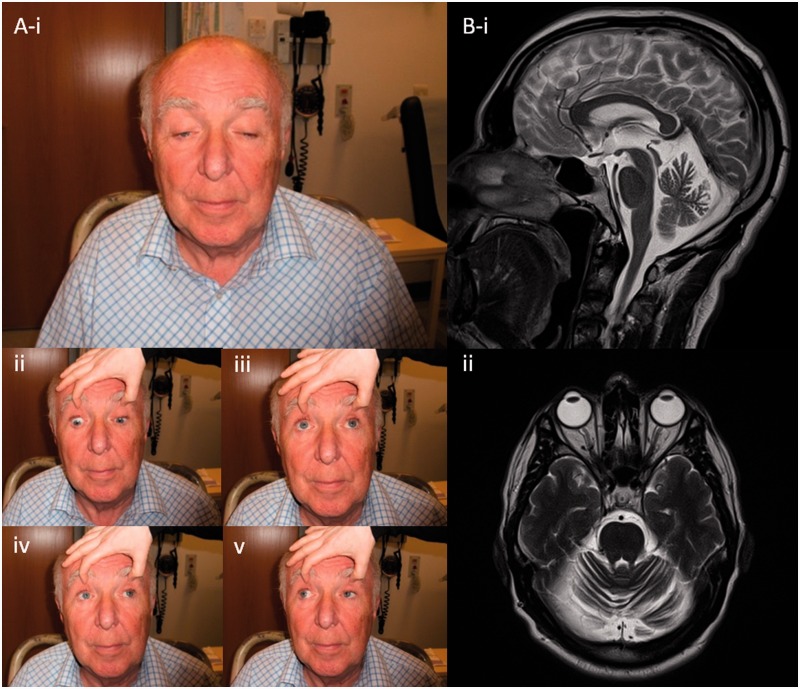
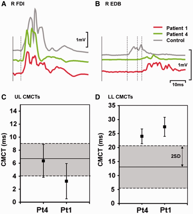
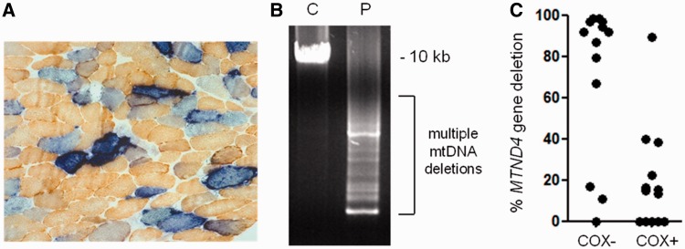
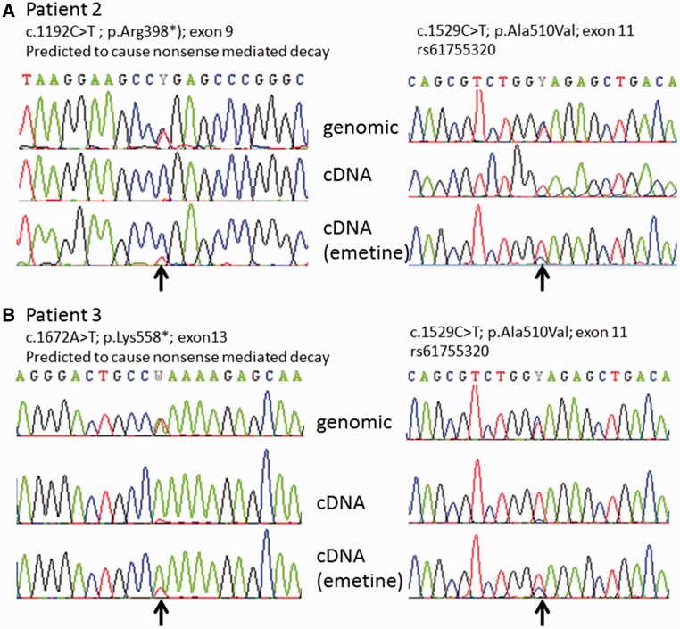
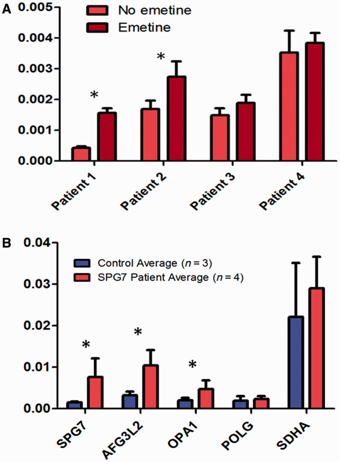
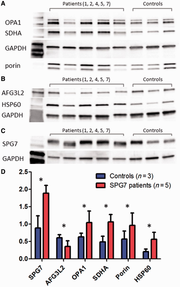
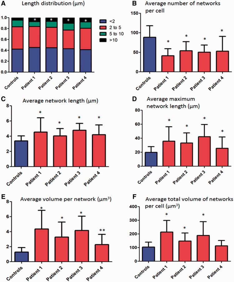
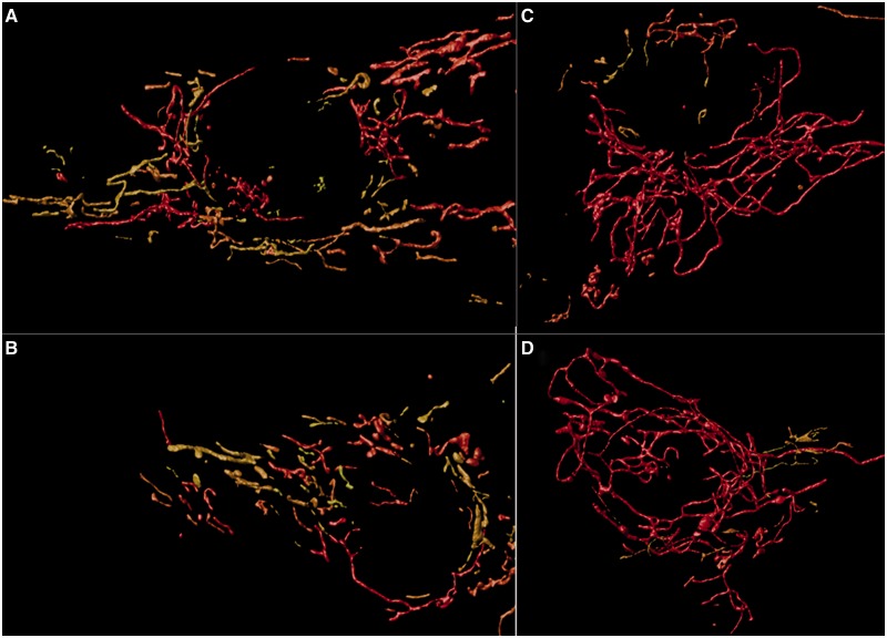
Despite being a canonical presenting feature of mitochondrial disease, the genetic basis of progressive external ophthalmoplegia remains unknown in a large proportion of patients. Here we show that mutations in SPG7 are a novel cause of progressive external ophthalmoplegia associated with multiple mitochondrial DNA deletions. After excluding known causes, whole exome sequencing, targeted Sanger sequencing and multiplex ligation-dependent probe amplification analysis were used to study 68 adult patients with progressive external ophthalmoplegia either with or without multiple mitochondrial DNA deletions in skeletal muscle. Nine patients (eight probands) were found to carry compound heterozygous SPG7 mutations, including three novel mutations: two missense mutations c.2221G>A; p.(Glu741Lys), c.2224G>A; p.(Asp742Asn), a truncating mutation c.861dupT; p.Asn288*, and seven previously reported mutations. We identified a further six patients with single heterozygous mutations in SPG7, including two further novel mutations: c.184-3C>T (predicted to remove a splice site before exon 2) and c.1067C>T; p.(Thr356Met). The clinical phenotype typically developed in mid-adult life with either progressive external ophthalmoplegia/ptosis and spastic ataxia, or a progressive ataxic disorder. Dysphagia and proximal myopathy were common, but urinary symptoms were rare, despite the spasticity. Functional studies included transcript analysis, proteomics, mitochondrial network analysis, single fibre mitochondrial DNA analysis and deep re-sequencing of mitochondrial DNA. SPG7 mutations caused increased mitochondrial biogenesis in patient muscle, and mitochondrial fusion in patient fibroblasts associated with the clonal expansion of mitochondrial DNA mutations. In conclusion, the SPG7 gene should be screened in patients in whom a disorder of mitochondrial DNA maintenance is suspected when spastic ataxia is prominent. The complex neurological phenotype is likely a result of the clonal expansion of secondary mitochondrial DNA mutations modulating the phenotype, driven by compensatory mitochondrial biogenesis.

Keywords: SPG7; chronic progressive external ophthalmoplegia; hereditary spastic paraplegia; mtDNA maintenance; paraplegin.


Figures

Figure 1
Figure 1
Clinical features. (A) Typical ophthalmological features of a patient with hereditary spastic paraplegia type 7 with PEO: marked ptosis is evident in primary gaze (i); extraocular motility in cardinal directions of gaze is mildly reduced, with restriction of upgaze most affected as the patient is asked to look down (ii); up (iii); left (iv); and right (v). (B) T2-weighted MRI images in demonstrating diffuse cerebellar volume loss, in sagittal (i); and transverse-axial (ii) planes.
Figure 2
Figure 2
Motor evoked potentials in Patients 1 and 4. Average (n = 10) rectified motor cortical evoked potentials (MEPs) recorded from (A) hand muscle, the right first dorsal interosseous (R FDI) and (B) foot muscle right extensor digitorum brevis (R EDB) in an age-matched male control (aged 64) shown in grey, Patient 1 (aged 65) in green and Patient 4 (aged 66) in red. Traces have been aligned after subtracting peripheral motor conduction times. Dashed lines indicate the onset of each MEP. Average central motor conduction times (mean ± 1 SD) for (C) right first dorsal interosseous and (D) right extensor digitorum brevis in the same patients. Average central motor conduction times (CMCTs) were calculated by subtracting the average peripheral motor conduction time (n = 10) from the average motor cortical evoked potential latency (n = 10), measured from unrectified EMG. The solid horizontal lines show the mean, dashed horizontal lines and grey shaded areas show 2 SD of the mean from published normal data (Eisen and Shtybel, 1990).
Figure 3
Figure 3
Characterization of mitochondrial DNA maintenance defect in Patient 1. (A) Sequential COX-SDH histochemistry demonstrates a mosaic distribution of COX-deficient muscle fibres (blue) amongst fibres exhibiting normal COX activity (brown), with significant evidence of mitochondrial proliferation as shown by enhanced SDH reactivity around the subsarcolemmal region of the muscle fibre (ragged-blue fibres). (B) Long range PCR amplification of muscle DNA across the major arc shows significant evidence of multiple mitochondrial DNA deletions. C = Control; P = patient. (C) Quantitative, single fibre real-time-PCR reveals the majority—but not all—of COX-deficient fibres contain high levels of a clonally-expanded mitochondrial DNA deletion involving the MTND4 gene, an observation which is consistent with multiple mitochondrial DNA deletions due to a disturbance of mitochondrial DNA maintenance (He et al., 2002).
Figure 4
Figure 4
Confirmatory complementary DNA sequencing in Patients 2 and 3. Arrows indicate the positions of the mutations. (A) In Patient 2, the c.1192C>T (p.Arg398*) mutation is predicted to cause nonsense mediated messenger RNA decay. At left the genomic sequence demonstrates this mutation to be present in the heterozygous state although in complementary DNA from fibroblasts it appears absent because it is degraded by nonsense mediated decay. The presence of this variant is partially restored in fibroblasts grown with emetine (an inhibitor of nonsense mediated decay). The second mutation in this patient [c.1529C>T; p.(Ala510Val)] is present at homozygous levels in complementary DNA indicating that it is on the opposite allele; as before the second allele is partially restored with emetine treatment. (B) Patient 3 has a c.1672A>T (p.Lys558*) mutation which similarly is predicted to cause nonsense mediated decay. The mutation is almost absent in complementary DNA but partially restored in cells grown with emetine. The second c.1529C>T; p.(Ala510Val) mutation is again shown to be present on the opposite allele.
Figure 5
Figure 5
Transcript level measurement with reverse transcription quantitative PCR. (A) SPG7 transcript analysis in Patients 1–4, using RNA from cultured fibroblasts. For Patients 1–3 who have one or more nonsense mutations in SPG7, treatment of fibroblasts with emetine (dark bars) (an inhibitor of nonsense-mediated messenger RNA decay) compared with normal conditions (light bars), resulted in significantly increased SPG7 messenger RNA in Patients 1 and 2 (*P < 0.05). Patient 4 who has 2 missense SPG7 mutations does not have alteration in SPG7 expression with emetine (nor do control; data not shown). (B) Transcript quantitation in complementary DNA derived from muscle, in Patients 1–4 (red bands), compared with three control muscle samples (blue bands). Levels of SPG7, AFG3L2, and OPA1 transcripts are elevated in patients compared with controls (*P < 0.02). Levels of POLG and SDHA did not differ significantly.
Figure 6
Figure 6
Western blot of muscle tissue. (A–C) Representative blots used in the quantification of protein expression of muscle tissue from five patients with compound heterozygous SPG7 mutations and three control subjects. Testing was performed in triplicate and quantification of aggregate mean with SD (normalized to GAPDH) are represented in D. Markers of mitochondrial mass, including SDHA, porin, and HSP60 are significantly increased in SPG7 patients compared with controls. SPG7 and OPA1 were also significantly elevated in SPG7 patients compared with controls. AGF3L2 was decreased compared with controls. All the above were statistically significant to *P < 0.01.
Figure 7
Figure 7
Mitochondrial network analysis. Mitochondrial network analysis was undertaken in fibroblasts (Patients 1–4) grown concurrently with identical medium and conditions. Error bars are SD. Controls are the aggregate results of four separate cell lines (50 cells each for total 200 cells). (A) The distribution of the network lengths is demonstrated; very long networks (>10 µm) were significantly more abundant in patients with compound heterozygous SPG7 mutations than controls. (B) The average number of total networks per cell was significantly lower in SPG7 patients. (C) The average length per mitochondrial network was increased in SPG7 patients, as was the average longest network per cell (D). The volume of individual mitochondrial networks was higher than controls per cell line (E) and the total volume of the mitochondrial network per cell was elevated (in all cell lines except Patient 4 which was not significant) (F). We suggest that these hyperfused mitochondrial networks may be a compensatory mechanism, and that the elevated total mitochondrial volume corresponds to the elevated mitochondrial mass observed in COX-deficient fibres in these patients. *P < 0.0001 and **P = 0.004.
Figure 8
Figure 8
Representative images from mitochondrial network analysis. Three-dimensional reconstruction of mitochondrial networks using Huygens Essentials software. Networks are colour-coded, in which short networks are yellow and longer networks are red. (A and B) Representative images of the networks from two separate control cell lines. (C and D) Representative images from cell lines derived from Patients 1 and 4, respectively. Qualitatively, one can observe that networks appear to be longer in the patient with compound heterozygous SPG7 mutations. Statistical analysis indicated that SPG7 patient cell networks were on average longer, with fewer networks but increased total volume of mitochondria.
Figure 9
Figure 9
Ultra-deep resequencing of mitochondrial DNA control region. Ultra-deep resequencing by synthesis (UDS) of skeletal muscle mitochondrial DNA. UDS (Roche 454 FLX Titanium) mitochondrial DNA hypervariable segment 2 (MT-HV2) amplicon. Comparison is made between a cloned segment (expected to be homoplasmic), with controls, and patients with genetically-confirmed mitochondrial DNA maintenance disorders due to recessive POLG, dominant OPA1, or recessive SPG7 mutations. The mutation burden in SPG7 patients was not statistically different from control subjects or OPA1 mutation carriers, but was significantly lower than POLG patients.

References

    1. Almajan ER, Richter R, Paeger L, Martinelli P, Barth E, Decker T, et al. AFG3L2 supports mitochondrial protein synthesis and purkinje cell survival. J Clin Invest. 2012;122:4048–58. - PMC - PubMed
    1. Arnoldi A, Tonelli A, Crippa F, Villani G, Pacelli C, Sironi M, et al. A clinical, genetic, and biochemical characterization of SPG7 mutations in a large cohort of patients with hereditary spastic paraplegia. Hum Mutat. 2008;29:522–31. - PubMed
    1. Baker MR, Fisher KM, Whittaker RG, Griffiths PG, Yu-Wai-Man P, et al. Subclinical multisystem neurologic disease in “pure” OPA1 autosomal dominant optic atrophy. Neurology. 2011;77:1309–12. - PMC - PubMed
    1. Blakely EL, Butterworth A, Hadden RD, Bodi I, He L, McFarland R, et al. MPV17 mutation causes neuropathy and leukoencephalopathy with multiple mtDNA deletions in muscle. Neuromuscul Disord. 2012;22:587–91. - PMC - PubMed
    1. Blakely E, He L, Gardner JL, Hudson G, Walter J, Hughes I, et al. Novel mutations in the TK2 gene associated with fatal mitochondrial DNA depletion myopathy. Neuromuscul Disord. 2008;18:557–60. - PubMed

Publication types

MeSH terms

Substances