Review
. 2014;90(4):119-29.
doi: 10.2183/pjab.90.119.

Ribosomal RNA gene repeats, their stability and cellular senescence

Affiliations
Review

Ribosomal RNA gene repeats, their stability and cellular senescence

Takehiko Kobayashi. Proc Jpn Acad Ser B Phys Biol Sci. 2014.

Abstract

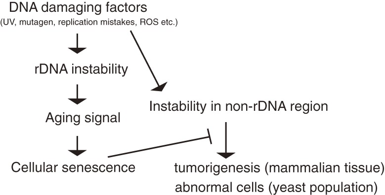
The ribosomal RNA gene (rDNA) repeats form a historically well-researched region in the chromosome. Their highly repetitive structure can be identified easily which has enabled studies on DNA replication, recombination, and transcription. The region is one of the most unstable regions in the genome because of deleterious recombination among the repeats. The ribosomal RNA gene repeats use a unique gene amplification system to restore the copy number after this has been reduced due to recombination. It has been shown that unstable features in the genome can accelerate cellular senescence that restricts the lifespan of a cell. Here, I will introduce a study by our group that shows how the stability of rDNA is maintained and affects lifespan. I propose that the ribosomal RNA gene repeats constitute a center from which the stability of the whole genome is regulated and the lifespan of the cell is controlled.


Figures

Figure 1.
Figure 1.
Structure of rDNA in bacteria, yeast and human cells. (A) In bacteria, Escherichia coli, 7 ribosomal RNA genes are mainly located in a region of the genome that contains an origin of replication. The direction of rRNA transcription (shown by green arrow heads) by RNA polymerase is the same as that of the replication forks (shown by red arrows). (B) In budding yeast (Saccharomyces cerevisiae), an rDNA repeating unit contains a gene for the 35S rRNA precursor that is processed into three mature rRNA species (18S, 5.8S, and 25S rRNA), an rARS (replication origin), a 5S rDNA, an E-pro (non-coding promoter) and an RFB (replication fork blocking site). Restriction by BglII (Bg) produces ∼4.6 kb rDNA fragments. (C) In a human cell, a unit contains an ori (replication origin), an RFB (replication fork blocking site, located in the Sal1 box), the gene for 47S rRNA which is processed into three mature rRNA species (18S, 5.8S, and 28S rRNA) and an NP (non-coding promoter). In yeast, the 5S ribosomal RNA genes localize to the intergenic spacer within the rDNA repeats while in a human cell, these genes are organized in independent repeats.
Figure 2.
Figure 2.
Detection of rDNA by electrophoresis. Yeast DNA (S. cerevisiae) digested by restriction enzyme BglII was analyzed by (A) conventional agarose gel electrophoresis and stained with ethidium bromide or by (B) two dimensional agarose gel electrophoresis that separates replication intermediates. After electrophoresis, southern hybridization was performed to detect the rDNA. (C) A cartoon to explain the pattern of (B). The intermediates (gray) with a branched structure migrate more slowly than linear molecules. In (A), as a size marker, lambda DNA digested by HindIII (M) is run along the digested yeast DNA (S) with the rDNA fragment indicated (arrow head).
Figure 3.
Figure 3.
Transcription of rDNA in budding yeast. Ongoing transcription of rDNA as visualized by electron microscopy (Miller spread)38) in cells from (A)(B) reduced rDNA strains (∼40 repeats) or (C) the wild type strain (∼150). The black and gray arrows indicate the direction of the transcription units in the active and inactive units, respectively. This picture is from French et al. (2003).14)
Figure 4.
Figure 4.
Inactive (silenced) units of rDNA provide “working space” for repair. Condensin associates with silenced units and connects sister-chromatids to facilitate recombinational repair. Lack of inactive units reduces the efficiency of repair and increases the sensitivity to DNA damage.
Figure 5.
Figure 5.
rDNA repeat number is unstable. (A) DNA damage may reduce the number of rDNA repeats. DNA damage that has occurred in an rDNA unit may be repaired by homologous recombination with another repeat (donor) in the same chromosome. In this case, units between the damage and donor may be lost by crossing-over when DNA is exchanged during recombination. (B) The number of rDNA repeats is always changing. Pulsed-field gel electrophoresis of eight colonies that originated from a single cell. The length of chromosome XII containing the rDNA is different in each colony because of the variation in the number of rDNA repeats. M is a DNA size maker (H. wingei chromosomes). This picture is from Kobayashi (2009).43)
Figure 6.
Figure 6.
Model of gene amplification in rDNA. (A) In normal situations (rDNA copy number is ∼150), the silencing protein, Sir2, represses E-pro (E-pro OFF), allowing the cohesin complex (dotted ellipse) to associate with the IGS. DSBs are repaired by equal sister chromatid recombination, with no change in the copy number. (B) In situations where copy number is reduced, Sir2 repression is removed and E-pro is activated (E-pro ON). The transcription displaces cohesin complex from the IGS and unequal sister chromatids can be used as templates for repair of DSBs. As the result, some units are replicated twice and rDNA copy number increases. The gray lines represent single chromatids (double-strand DNA).
Figure 7.
Figure 7.
“Budding” in yeast. (A) A bud on the mother cell grows into a daughter cell. White rings on the surface of the mother cell are scars that are traces of budding. The smaller daughter cell is separated from the mother cell at the end of M phase. (B) A cartoon to explain (A).
Figure 8.
Figure 8.
The rDNA theory for aging. The rDNA region is sensitive to DNA damage by internal and external causes. Through cell divisions, the rDNA becomes more unstable than other parts of the genome and produces an aging signal that tells the cell to die. This precaution prevents the appearance of abnormal cells in a population or a tissue.

References

    1. Ellwood M., Nomura M. (1982) Chromosomal locations of the genes for rRNA in Escherichia coli K-12. J. Bacteriol. 149, 458–468 - PMC - PubMed
    1. Nomura M., Morgan E.A., Jaskunas S.R. (1977) Genetics of bacterial ribosomes. Annu. Rev. Genet. 11, 297–347 - PubMed
    1. Warner J.R. (1999) The economics of ribosome biosynthesis in yeast. Trends Biochem. Sci. 24, 437–440 - PubMed
    1. Hartwell, L.H. (2008) Genetics: From Genes to Genomes, 3rd Edition. McGraw-Hill, U.S.A.
    1. Brewer B.J., Fangman W.L. (1988) Mapping replication origins in yeast chromosomes. Bioessays 13, 317–322 - PubMed

Publication types