

Redefining the PF06864 Pfam family based on Burkholderia pseudomallei PilO2(Bp) S-SAD crystal structure
- PMID: 24728008
- PMCID: PMC3984277
- DOI: 10.1371/journal.pone.0094981
Redefining the PF06864 Pfam family based on Burkholderia pseudomallei PilO2(Bp) S-SAD crystal structure
Abstract
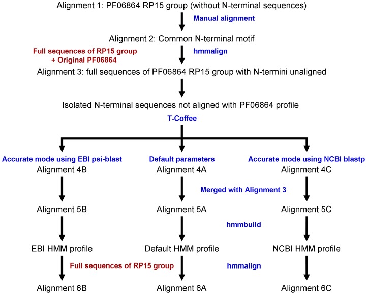
Type IV pili are surface-exposed filaments and bacterial virulence factors, represented by the Tfpa and Tfpb types, which assemble via specific machineries. The Tfpb group is further divided into seven variants, linked to heterogeneity in the assembly machineries. Here we focus on PilO2(Bp), a protein component of the Tfpb R64 thin pilus variant assembly machinery from the pathogen Burkholderia pseudomallei. PilO2(Bp) belongs to the PF06864 Pfam family, for which an improved definition is presented based on newly derived Hidden Markov Model (HMM) profiles. The 3D structure of the N-terminal domain of PilO2(Bp) (N-PilO2(Bp)), here reported, is the first structural representative of the PF06864 family. N-PilO2(Bp) presents an actin-like ATPase fold that is shown to be present in BfpC, a different variant assembly protein; the new HMM profiles classify BfpC as a PF06864 member. Our results provide structural insight into the PF06864 family and on the Type IV pili assembly machinery.
Conflict of interest statement
Figures


Similar articles
-
Production and purification of Burkholderia pseudomallei BipD protein.Southeast Asian J Trop Med Public Health. 2008 Jan;39(1):109-14. Southeast Asian J Trop Med Public Health. 2008. PMID: 18567449
-
A novel FK-506-binding-like protein that lacks peptidyl-prolyl isomerase activity is involved in intracellular infection and in vivo virulence of Burkholderia pseudomallei.Microbiology (Reading). 2011 Sep;157(Pt 9):2629-2638. doi: 10.1099/mic.0.049163-0. Epub 2011 Jun 16. Microbiology (Reading). 2011. PMID: 21680634
-
A second type III secretion system in Burkholderia pseudomallei: who is the real culprit?Microbiology (Reading). 2001 Dec;147(Pt 12):3197-9. doi: 10.1099/00221287-147-12-3197. Microbiology (Reading). 2001. PMID: 11739751 No abstract available.
-
A novel contact-independent T6SS that maintains redox homeostasis via Zn2+ and Mn2+ acquisition is conserved in the Burkholderia pseudomallei complex.Microbiol Res. 2019 Sep;226:48-54. doi: 10.1016/j.micres.2019.05.007. Epub 2019 May 31. Microbiol Res. 2019. PMID: 31284944 Review.
-
Molecular insights into Burkholderia pseudomallei and Burkholderia mallei pathogenesis.Annu Rev Microbiol. 2010;64:495-517. doi: 10.1146/annurev.micro.112408.134030. Annu Rev Microbiol. 2010. PMID: 20528691 Review.
Cited by
-
Crystallization and preliminary crystallographic studies of the hypothetical protein BPSL1038 from Burkholderia pseudomallei.Acta Crystallogr F Struct Biol Commun. 2014 Dec 1;70(Pt 12):1697-700. doi: 10.1107/S2053230X14025278. Epub 2014 Nov 28. Acta Crystallogr F Struct Biol Commun. 2014. PMID: 25484229 Free PMC article.
-
Boosting of post-exposure human T-cell and B-cell recall responses in vivo by Burkholderia pseudomallei-related proteins.Immunology. 2017 May;151(1):98-109. doi: 10.1111/imm.12709. Epub 2017 Feb 9. Immunology. 2017. PMID: 28066900 Free PMC article.
-
Melioidosis: molecular aspects of pathogenesis.Expert Rev Anti Infect Ther. 2014 Dec;12(12):1487-99. doi: 10.1586/14787210.2014.970634. Epub 2014 Oct 14. Expert Rev Anti Infect Ther. 2014. PMID: 25312349 Free PMC article. Review.
References
Publication types
MeSH terms
Substances
LinkOut - more resources
Full Text Sources
Other Literature Sources
