. 2014 Apr 11;3(5):353-61.
doi: 10.1242/bio.20147708.

Colchicine-induced degeneration of the micronucleus during conjugation in Tetrahymena

Affiliations

Colchicine-induced degeneration of the micronucleus during conjugation in Tetrahymena

Pin-Fang Chen et al. Biol Open. .

Abstract

One of the most dramatic examples of nuclear morphogenesis occurs during conjugation in Tetrahymena when the micronucleus elongates to a size longer than the cell itself. After contraction to a spherical shape, the nucleus moves directly to chromosome separation in the first meiotic division. Here we investigate the consequences of interrupting the elongation process. Colchicine, a microtubule inhibitor, caused retraction of elongated structures. With time, cells began to lose their micronuclei, and by five hours more than half of the paired cells had at least one cell missing a micronucleus. After reversing the colchicine block, existing micronuclei did not undergo elongation again, nor did meiosis occur. These observations indicate that micronuclear elongation is critical to subsequent meiotic division. Further, nuclear elimination occurs, which could be due to meiotic failure or possibly a problem downstream from meiosis. An analysis of the process of colchicine-induced micronuclear degeneration indicated that it was regulated by a caspase-dependent mechanism, characteristic of apoptosis, and then resorbed by a lysosome-dependent autophagic mechanism. Amicronucleate cells failed to grow when returned to nutrient medium, likely because of a lesion in the post-conjugation reconstruction of a functioning oral apparatus. The ease by which a large number of nuclei are induced to "self-destruct" may make this system useful in investigating the link between colchicine treatment and nuclear death in Tetrahymena, and in investigating how nuclear death could be regulated in living cells more generally. Finally, we note that this phenomenon might relate to the evolution of amicronucleate species of Tetrahymena.

Keywords: Apoptosis; Autophagy; Ciliate; Meiosis; Micronucleus; Nuclear morphogenesis; Tetrahymena.


Conflict of interest statement

Competing interests: The authors have no competing interests to declare.

Figures

Fig. 1.
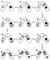
Fig. 1.. Diagram of the nuclear stages that occur during conjugation of mating types of Tetrahymena.
The rounded micronucleus moves from its pocket in the macronucleus (a), elongates (b), then contracts and undergoes 2 meiotic divisions forming four haploid products (c). Three haploid nuclei degrade (d) and the remaining haploid nucleus divides by mitosis to form two gametic pronuclei (e). After reciprocal exchange (f), fertilization occurs forming a diploid zygote nucleus (g). The zygote nucleus divides twice mitotically to form four nuclei (h), two of which enlarge to form developing macronuclei and two of which form micronuclei, while the old macronucleus condenses and moves to the posterior end of the cell (k,m). The old macronucleus is then eliminated (n,p).
Fig. 2.
Fig. 2.. DAPI-stained cells during conjugation.
Uninterrupted micronuclear elongation, and elongation reversed with colchicine. Shortly after cell pairing the micronucleus (arrows) migrates slightly from its position in a macronuclear pocket to a cytoplasmic position near the macronucleus (a). It then elongates and curls around the macronucleus (b). Elongated nuclei collapse (arrows) in response to colchicine (c) and may disappear from one (d) or both cells (not shown). Scale bars: 5.7 µm (a), 10 µm (b), 8 µm (c), 3.6 µm (d).
Fig. 3.
Fig. 3.. Cells were exposed to 5 mg/ml colchicine at 2½ hours after mixing mating types.
Samples were then fixed at hourly time points. Cells in pairs were scored for the presence or absence of a visible micronucleus. These data (open circles) show that the disappearance of the micronucleus increases with time. At 5 hours after adding colchicine micronuclei have been lost in more than half of all individual cells in pairs. Control cells also show some loss of micronuclei (marked ‘x’), which could reflect a spontaneous micronuclear loss, or could indicate error in this visual assay, or both. For this and other graphs, experiments were done in triplicate. Data points show averages, and bars represent range in variation.
Fig. 4.
Fig. 4.. Bar graph showing the effect of zVAD-fmk, a caspase inhibitor, on colchicine-induced micronuclear degeneration.
With colchicine alone (5 mg/ml, 2.5–7.5 hours after mixing mating types) about 50 percent of cells in pairs lose micronuclei. As in Fig. 3, even control cells, with no colchicine added, show some micronuclear loss. However, when zVAD is added together with colchicine, micronuclear loss is almost completely inhibited.
Fig. 5.
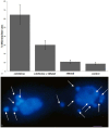
Fig. 5.. The effect of 3 methyladenine, an inhibitor of autophagy.
(a) Bar graph showing the effect of 3 methyladenine, an inhibitor of autophagy, on CIMD. In this set of experiments, about 60% of cells in pairs lost their micronuclei with colchicine (5 mg/ml, 2.5–7.5 hours after mixing mating types). However, when 3 methyladenine was added together with colchicine, micronuclear loss was reduced by about half. Control cells without colchicine, and cells with 3 methyladenine, also show some micronuclear loss. (b) DAPI stained cells after prolonged exposure to 3 methyladenine. 3 Methyladenine (15 mM) was added at 4 hours after mixing mating types, and assayed at 12 hours. In addition to two large macronuclei, which did not condense, nor were they resorbed, 14 smaller nuclei are visible in two cells of a pair (arrows). (Nuclei can shift from one cell to another when large openings in the conjugation junction occur.) This is the predicted result if developmentally programmed resorption of nuclei is prevented. Scale bar: 5 µm.
Fig. 6.
Fig. 6.. Line graph showing the effect of cytochalasin D, an actin inhibitor, on colchicine-induced micronuclear elimination.
Cytochalasin D was solubilized and diluted to different concentrations in 2% DMSO. The graph shows the extent of micronuclear loss with colchicine alone (5 mg/ml, 2.5–7.5 hours after mixing mating types), 2% DMSO alone, and colchicine with cytochalasin in DMSO, at increasing concentrations. The data show that cytochalasin D blocks colchicine-induced micronuclear loss in a concentration-dependent manner. At 100 µg/ml, cytochalasin D almost completely eliminates the effect of colchicine on micronuclear loss. A measurable loss of micronuclei occurs with DMSO alone.
Fig. 7.
Fig. 7.. Live pairs double stained with Hoechst 33342 and acridine orange (AO) 3 hours after exposure to colchicine.
In all four cells in the two pairs shown in panels a and b, the micronucleus has collapsed. In one cell it is large and blue while in three of the four cells the micronucleus is diminished in size, clearly being resorbed, and is a yellow–green color (arrows), a result of the combination of orange from AO (acidic vesicle), and blue for the Hoechst 33342 (DNA). These data indicate that CIMD occurs in an acidic environment, suggestive of lysosome-dependent autophagy. Panel c shows an enlargement of a portion of panel a as indicated by the dashed-line boxes; the differences in size and staining color of the two types of micronuclei are quite evident. Scale bars: 13.3 µm (a,b), 3.75 µm (c).
Fig. 8.
Fig. 8.. Texas Red Dextran moves from lysosomes into degenerating nuclei.
Both cells in the pair in panel a show a single parental macronucleus in the process of degradation. The macronucleus in the left cell is smaller (asterisk), indicating that it is further along in resorption. One can see orange stain in the macronucleus indicating that TRD loaded into lysosome was transferred to the macronucleus, as predicted for autophagy. In panels b and c, cells were incubated in Texas Red Dextran for 1 hour before mixing mating types, and were then exposed to colchicine at 2.5 hours after mixing mating types. Cells were fixed 3 hours later. Concentrated foci of TRD are seen in the posterior ends of a pair of cells in panel b. The same pair is seen in panel c stained with DAPI. The long arrow points to a micronucleus, which cannot be seen in panel b where it is masked by the TRD staining. The arrowhead in panel c points to an aggregate of TRD-containing lysosomes; no micronucleus is visible in the same cell in panel c. Presumably, a micronucleus had been there but is completely resorbed. Panel d is a photographically superimposed image composed of panels b and c, using Photoshop. The small silvery micronucleus in the cell on the right (arrow) is embedded in an aggregate of red TRD-containing lysosomes; in panel b the micronucleus is indistinguishable from the lysosomes indicating that it is loaded with TRD transferred from lysosomes. Scale bars: 7.5 µm (a), 7.2 µm (b–d).

Similar articles

Cited by

References

    1. Akematsu T., Endoh H. (2010). Role of apoptosis-inducing factor (AIF) in programmed nuclear death during conjugation in Tetrahymena thermophila. BMC Cell Biol. 11, 13 10.1186/1471-2121-11-13 - DOI - PMC - PubMed
    1. Akematsu T., Pearlman R. E., Endoh H. (2010). Gigantic macroautophagy in programmed nuclear death of Tetrahymena thermophila. Autophagy 6, 901–911 10.4161/auto.6.7.13287 - DOI - PMC - PubMed
    1. Allen D. L., Linderman J. K., Roy R. R., Bigbee A. J., Grindeland R. E., Mukku V., Edgerton V. R. (1997). Apoptosis: a mechanism contributing to remodeling of skeletal muscle in response to hindlimb unweighting. Am. J. Physiol. 273, C579–C587. - PubMed
    1. Bassnett S. (2009). On the mechanism of organelle degradation in the vertebrate lens. Exp. Eye Res. 88, 133–139 10.1016/j.exer.2008.08.017 - DOI - PMC - PubMed
    1. Bruusgaard J. C., Egner I. M., Larsen T. K., Dupre-Aucouturier S., Desplanches D., Gundersen K. (2012). No change in myonuclear number during muscle unloading and reloading. J. Appl. Physiol. 113, 290–296 10.1152/japplphysiol.00436.2012 - DOI - PubMed

LinkOut - more resources