

Cone photoreceptor definition on adaptive optics retinal imaging
- PMID: 24729030
- PMCID: PMC4112439
- DOI: 10.1136/bjophthalmol-2013-304615
Cone photoreceptor definition on adaptive optics retinal imaging
Abstract
Aims: To quantitatively analyse cone photoreceptor matrices on images captured on an adaptive optics (AO) camera and assess their correlation to well-established parameters in the retinal histology literature.
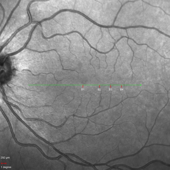
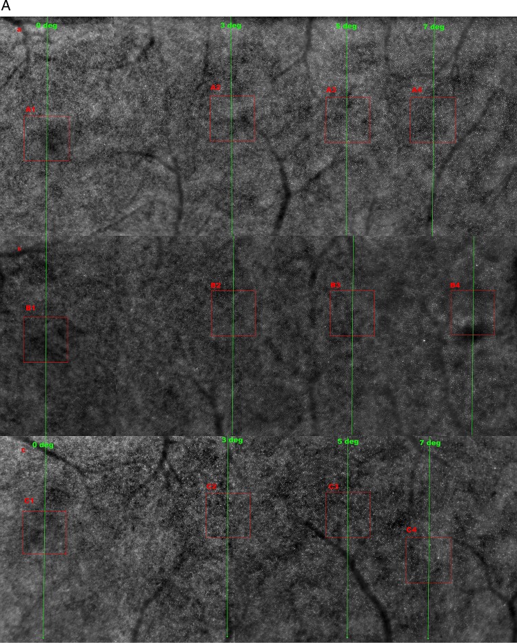
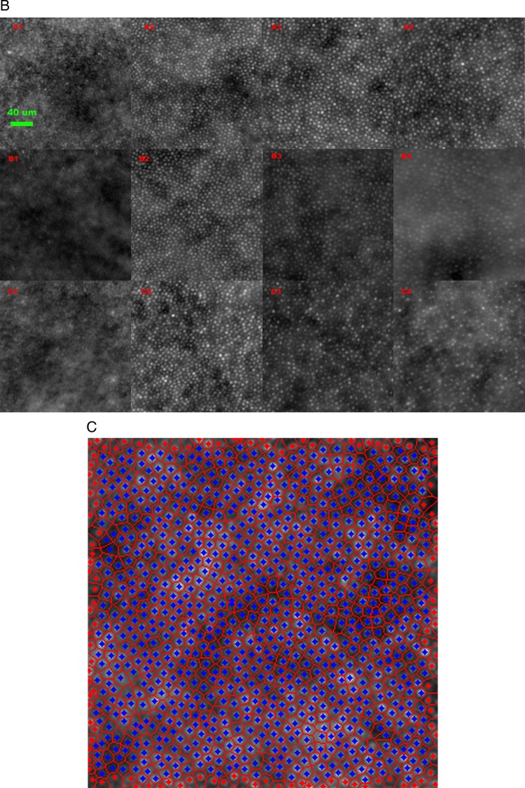
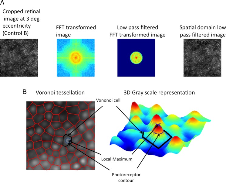
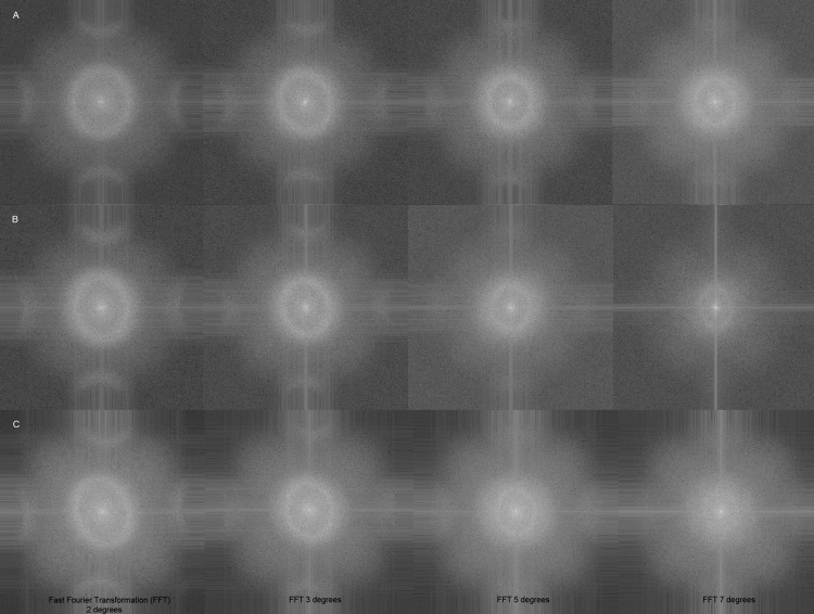
Methods: High resolution retinal images were acquired from 10 healthy subjects, aged 20-35 years old, using an AO camera (rtx1, Imagine Eyes, France). Left eye images were captured at 5° of retinal eccentricity, temporal to the fovea for consistency. In three subjects, images were also acquired at 0, 2, 3, 5 and 7° retinal eccentricities. Cone photoreceptor density was calculated following manual and automated counting. Inter-photoreceptor distance was also calculated. Voronoi domain and power spectrum analyses were performed for all images.
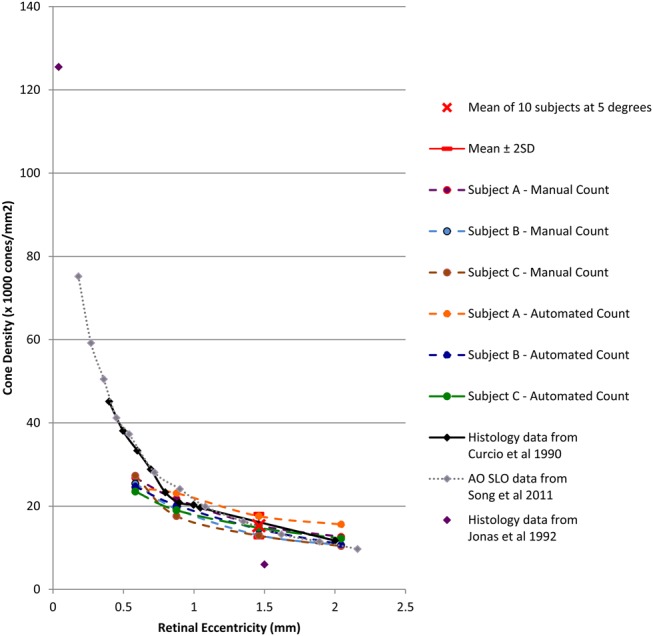
Results: At 5° eccentricity, the cone density (cones/mm(2) mean±SD) was 15.3±1.4×10(3) (automated) and 13.9±1.0×10(3) (manual) and the mean inter-photoreceptor distance was 8.6±0.4 μm. Cone density decreased and inter-photoreceptor distance increased with increasing retinal eccentricity from 2 to 7°. A regular hexagonal cone photoreceptor mosaic pattern was seen at 2, 3 and 5° of retinal eccentricity.
Conclusions: Imaging data acquired from the AO camera match cone density, intercone distance and show the known features of cone photoreceptor distribution in the pericentral retina as reported by histology, namely, decreasing density values from 2 to 7° of eccentricity and the hexagonal packing arrangement. This confirms that AO flood imaging provides reliable estimates of pericentral cone photoreceptor distribution in normal subjects.
Keywords: Adaptive Optics; Cone Photoreceptor; Retinal Imaging.
Published by the BMJ Publishing Group Limited. For permission to use (where not already granted under a licence) please go to http://group.bmj.com/group/rights-licensing/permissions.
Figures
Similar articles
-
Comparing Parafoveal Cone Photoreceptor Mosaic Metrics in Younger and Older Age Groups Using an Adaptive Optics Retinal Camera.Ophthalmic Surg Lasers Imaging Retina. 2017 Jan 1;48(1):45-50. doi: 10.3928/23258160-20161219-06. Ophthalmic Surg Lasers Imaging Retina. 2017. PMID: 28060393
-
Technical factors influencing cone packing density estimates in adaptive optics flood illuminated retinal images.PLoS One. 2014 Sep 9;9(9):e107402. doi: 10.1371/journal.pone.0107402. eCollection 2014. PLoS One. 2014. PMID: 25203681 Free PMC article.
-
Eccentricity dependent changes of density, spacing and packing arrangement of parafoveal cones.Ophthalmic Physiol Opt. 2013 Jul;33(4):516-26. doi: 10.1111/opo.12053. Epub 2013 Mar 29. Ophthalmic Physiol Opt. 2013. PMID: 23550537
-
Adaptive optics ophthalmoscopy.J Refract Surg. 2000 Sep-Oct;16(5):S602-7. doi: 10.3928/1081-597X-20000901-23. J Refract Surg. 2000. PMID: 11019882 Review.
-
Photoreceptor-Based Biomarkers in AOSLO Retinal Imaging.Invest Ophthalmol Vis Sci. 2017 May 1;58(6):BIO255-BIO267. doi: 10.1167/iovs.17-21868. Invest Ophthalmol Vis Sci. 2017. PMID: 28873135 Free PMC article. Review.
Cited by
-
Multilayer Retinal Correspondence of the Structural and Vascular Anomalies in Eyes With Early Macular Telangiectasia Type 2.Invest Ophthalmol Vis Sci. 2024 Sep 3;65(11):24. doi: 10.1167/iovs.65.11.24. Invest Ophthalmol Vis Sci. 2024. PMID: 39283616 Free PMC article.
-
Modeling Human Macular Cone Photoreceptor Spatial Distribution.Invest Ophthalmol Vis Sci. 2024 Jul 1;65(8):14. doi: 10.1167/iovs.65.8.14. Invest Ophthalmol Vis Sci. 2024. PMID: 38975943 Free PMC article.
-
Use of focus measure operators for characterization of flood illumination adaptive optics ophthalmoscopy image quality.Biomed Opt Express. 2018 Jan 18;9(2):679-693. doi: 10.1364/BOE.9.000679. eCollection 2018 Feb 1. Biomed Opt Express. 2018. PMID: 29552404 Free PMC article.
-
Quality improvement of adaptive optics retinal images using conditional adversarial networks.Biomed Opt Express. 2020 Jan 14;11(2):831-849. doi: 10.1364/BOE.380224. eCollection 2020 Feb 1. Biomed Opt Express. 2020. PMID: 32133226 Free PMC article.
-
Chromatic Full-Field Stimulus Thresholds in Patients with Treatment-Naive Age-Related Macular Degeneration.Clin Ophthalmol. 2022 Jan 29;16:223-229. doi: 10.2147/OPTH.S346291. eCollection 2022. Clin Ophthalmol. 2022. PMID: 35125864 Free PMC article.
References
-
- Liang J, Williams DR, Miller DT. Supernormal vision and high-resolution retinal imaging through adaptive optics. J Opt Soc Am A Opt Image Sci Vis 1997;14:2884–92 - PubMed
-
- Roorda A, Romero-Borja F, Donnelly Iii W, et al. Adaptive optics scanning laser ophthalmoscopy. Opt Expr 2002;10:405–12 - PubMed
-
- Roorda A, Williams DR. The arrangement of the three cone classes in the living human eye. Nature 1999;397:520–2 - PubMed
-
- Miller DT, Williams DR, Morris GM, et al. Images of cone photoreceptors in the living human eye. Vision Res 1996;36:1067–79 - PubMed
Publication types
MeSH terms
Grants and funding
LinkOut - more resources
Full Text Sources
Other Literature Sources