

Nature-inspired nanoformulations for contrast-enhanced in vivo MR imaging of macrophages
- PMID: 24729189
- PMCID: PMC4197124
- DOI: 10.1002/cmmi.1587
Nature-inspired nanoformulations for contrast-enhanced in vivo MR imaging of macrophages
Abstract
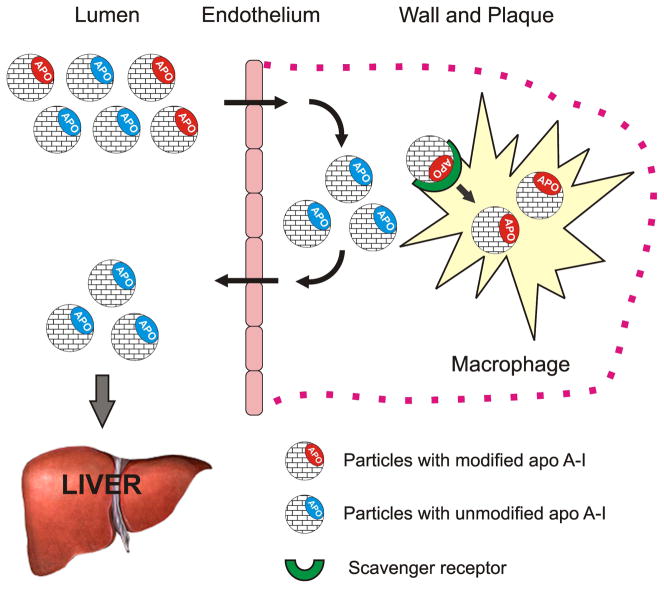
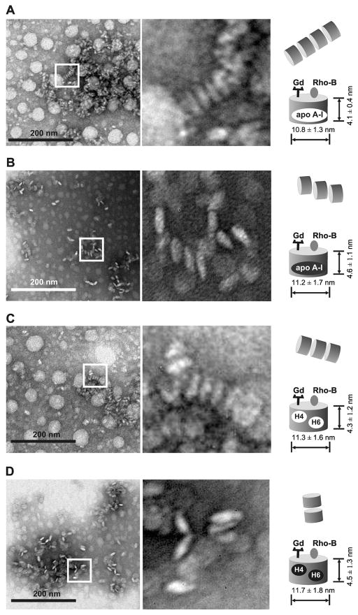
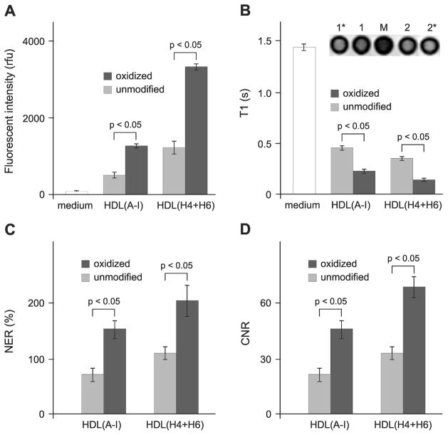
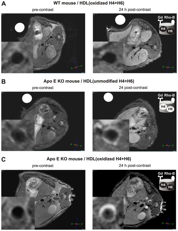
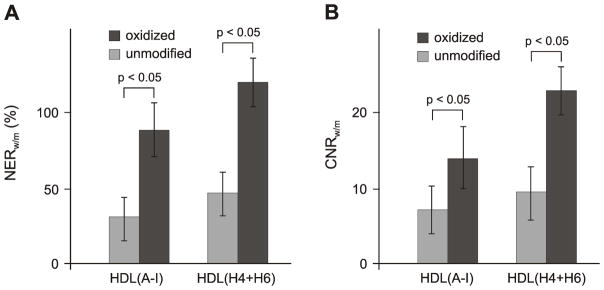
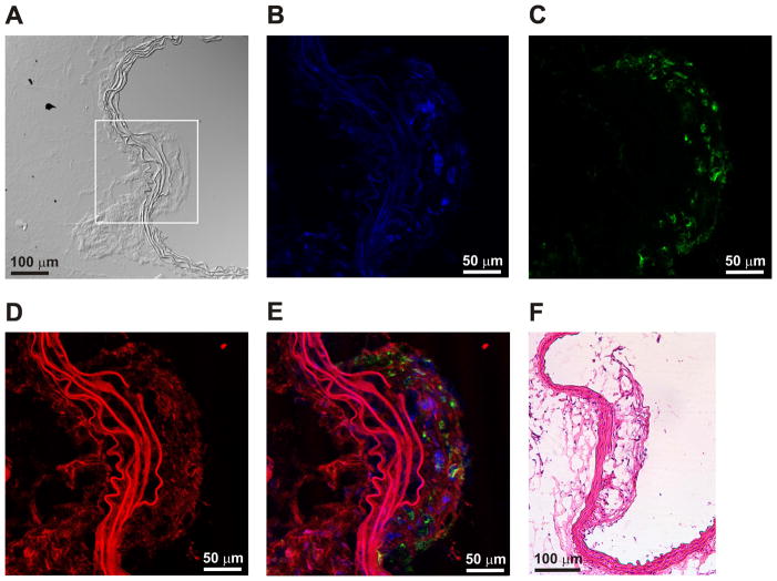
Magnetic resonance imaging (MRI) of macrophages in atherosclerosis requires the use of contrast-enhancing agents. Reconstituted lipoprotein particles that mimic native high-density lipoproteins (HDL) are a versatile delivery platform for Gd-based contrast agents (GBCA) but require targeting moieties to direct the particles to macrophages. In this study, a naturally occurring methionine oxidation in the major HDL protein, apolipoprotein (apo) A-I, was exploited as a novel way to target HDL to macrophages. We also tested if fully functional GBCA-HDL can be generated using synthetic apo A-I peptides. The fluorescence and MRI studies reveal that specific oxidation of apo A-I or its peptides increases the in vitro macrophage uptake of GBCA-HDL by 2-3 times. The in vivo imaging studies using an apo E-deficient mouse model of atherosclerosis and a 3.0 T MRI system demonstrate that this modification significantly improves atherosclerotic plaque detection using GBCA-HDL. At 24 h post-injection of 0.05 mmol Gd kg(-1) GBCA-HDL containing oxidized apo A-I or its peptides, the atherosclerotic wall/muscle normalized enhancement ratios were 90 and 120%, respectively, while those of GBCA-HDL containing their unmodified counterparts were 35 and 45%, respectively. Confocal fluorescence microscopy confirms the accumulation of GBCA-HDL containing oxidized apo A-I or its peptides in intraplaque macrophages. Together, the results of this study confirm the hypothesis that specific oxidation of apo A-I targets GBCA-HDL to macrophages in vitro and in vivo. Furthermore, our observation that synthetic peptides can functionally replace the native apo A-I protein in HDL further encourages the development of these contrast agents for macrophage imaging.
Keywords: apolipoprotein A-I; atherosclerosis; biomimetic peptide; gadolinium; lipoprotein nanoparticle; macrophage; magnetic resonance imaging; oxidation; vulnerable plaque.
Copyright © 2014 John Wiley & Sons, Ltd.
Figures
Similar articles
-
Diagnostic Magnetic Resonance Imaging of Atherosclerosis in Apolipoprotein E Knockout Mouse Model Using Macrophage-Targeted Gadolinium-Containing Synthetic Lipopeptide Nanoparticles.PLoS One. 2015 Nov 16;10(11):e0143453. doi: 10.1371/journal.pone.0143453. eCollection 2015. PLoS One. 2015. PMID: 26569115 Free PMC article.
-
Incorporation of an apoE-derived lipopeptide in high-density lipoprotein MRI contrast agents for enhanced imaging of macrophages in atherosclerosis.Contrast Media Mol Imaging. 2008 Nov-Dec;3(6):233-42. doi: 10.1002/cmmi.257. Contrast Media Mol Imaging. 2008. PMID: 19072768
-
Collagen-specific peptide conjugated HDL nanoparticles as MRI contrast agent to evaluate compositional changes in atherosclerotic plaque regression.JACC Cardiovasc Imaging. 2013 Mar;6(3):373-84. doi: 10.1016/j.jcmg.2012.06.016. Epub 2013 Feb 20. JACC Cardiovasc Imaging. 2013. PMID: 23433925 Free PMC article.
-
High-density lipoprotein-based contrast agents for multimodal imaging of atherosclerosis.Arterioscler Thromb Vasc Biol. 2010 Feb;30(2):169-76. doi: 10.1161/ATVBAHA.108.179275. Epub 2009 Oct 8. Arterioscler Thromb Vasc Biol. 2010. PMID: 19815819 Free PMC article. Review.
-
Histological validation of iron-oxide and gadolinium based MRI contrast agents in experimental atherosclerosis: the do's and don't's.Atherosclerosis. 2012 Dec;225(2):274-80. doi: 10.1016/j.atherosclerosis.2012.07.028. Epub 2012 Jul 27. Atherosclerosis. 2012. PMID: 22882907 Review.
Cited by
-
SARS Coronavirus Fusion Peptide-Derived Sequence Suppresses Collagen-Induced Arthritis in DBA/1J Mice.Sci Rep. 2016 Jun 28;6:28672. doi: 10.1038/srep28672. Sci Rep. 2016. PMID: 27349522 Free PMC article.
-
Inhibition of Triggering Receptor Expressed on Myeloid Cells 1 Ameliorates Inflammation and Macrophage and Neutrophil Activation in Alcoholic Liver Disease in Mice.Hepatol Commun. 2018 Oct 29;3(1):99-115. doi: 10.1002/hep4.1269. eCollection 2019 Jan. Hepatol Commun. 2018. PMID: 30619998 Free PMC article.
-
Molecular and Nonmolecular Imaging of Macrophages in Atherosclerosis.Front Cardiovasc Med. 2021 May 19;8:670639. doi: 10.3389/fcvm.2021.670639. eCollection 2021. Front Cardiovasc Med. 2021. PMID: 34095259 Free PMC article. Review.
-
Nanoparticle theranostics in cardiovascular inflammation.Semin Immunol. 2021 Aug;56:101536. doi: 10.1016/j.smim.2021.101536. Epub 2021 Nov 30. Semin Immunol. 2021. PMID: 34862118 Free PMC article. Review.
-
Inhibition of TREM-2 Markedly Suppresses Joint Inflammation and Damage in Experimental Arthritis.Int J Mol Sci. 2022 Aug 9;23(16):8857. doi: 10.3390/ijms23168857. Int J Mol Sci. 2022. PMID: 36012120 Free PMC article.
References
-
- Michaud CM, Murray CJ, Bloom BR. Burden of disease--implications for future research. Jama. 2001;285(5):535–539. - PubMed
-
- Shah PK. Pathophysiology of coronary thrombosis: role of plaque rupture and plaque erosion. Prog Cardiovasc Dis. 2002;44(5):357–368. - PubMed
-
- Bhatia V, Bhatia R, Dhindsa S, Virk A. Vulnerable plaques, inflammation and newer imaging modalities. J Postgrad Med. 2003;49(4):361–368. - PubMed
-
- Sharif F, Murphy RT. Current status of vulnerable plaque detection. Catheter Cardiovasc Interv. 2010;75(1):135–144. - PubMed
-
- Schaar JA, Mastik F, Regar E, den Uil CA, Gijsen FJ, Wentzel JJ, Serruys PW, van der Stehen AF. Current diagnostic modalities for vulnerable plaque detection. Curr Pharm Des. 2007;13(10):995–1001. - PubMed
Publication types
MeSH terms
Substances
Grants and funding
LinkOut - more resources
Full Text Sources
Other Literature Sources
Medical
