

Facile method for the site-specific, covalent attachment of full-length IgG onto nanoparticles
- PMID: 24729432
- PMCID: PMC4142076
- DOI: 10.1002/smll.201303629
Facile method for the site-specific, covalent attachment of full-length IgG onto nanoparticles
Abstract
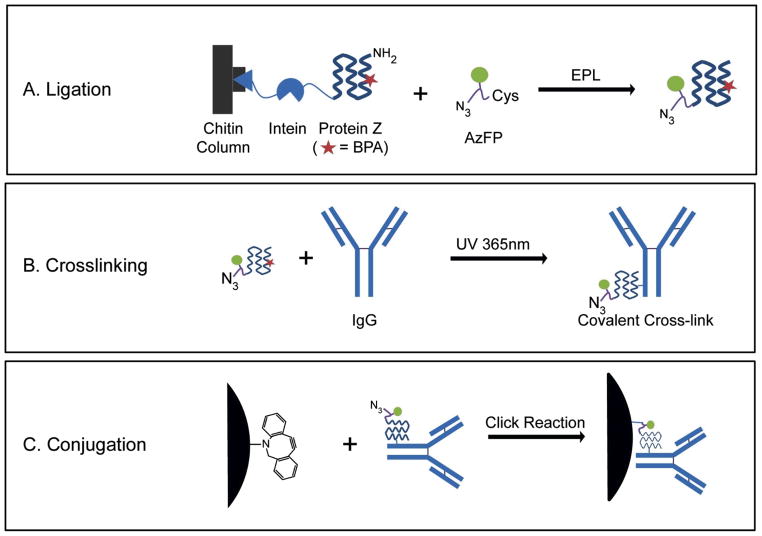
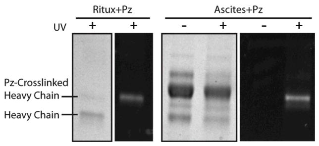
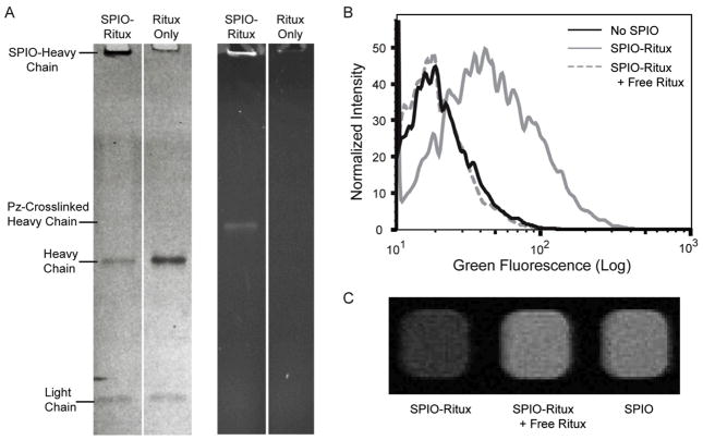
Antibodies, most commonly IgGs, have been widely used as targeting ligands in research and therapeutic applications due to their wide array of targets, high specificity and proven efficacy. Many of these applications require antibodies to be conjugated onto surfaces (e.g. nanoparticles and microplates); however, most conventional bioconjugation techniques exhibit low crosslinking efficiencies, reduced functionality due to non-site-specific labeling and random surface orientation, and/or require protein engineering (e.g. cysteine handles), which can be technically challenging. To overcome these limitations, we have recombinantly expressed Protein Z, which binds the Fc region of IgG, with an UV active non-natural amino acid benzoylphenyalanine (BPA) within its binding domain. Upon exposure to long wavelength UV light, the BPA is activated and forms a covalent link between the Protein Z and the bound Fc region of IgG. This technology was combined with expressed protein ligation (EPL), which allowed for the introduction of a fluorophore and click chemistry-compatible azide group onto the C-terminus of Protein Z during the recombinant protein purification step. This enabled the crosslinked-Protein Z-IgG complexes to be efficiently and site-specifically attached to aza-dibenzocyclooctyne-modified nanoparticles, via copper-free click chemistry.
Keywords: antibody; click chemistry; conjugation; nanoparticle; site-specific.
© 2014 WILEY-VCH Verlag GmbH & Co. KGaA, Weinheim.
Figures
Similar articles
-
Optimization of photoactive protein Z for fast and efficient site-specific conjugation of native IgG.Bioconjug Chem. 2014 Sep 17;25(9):1709-19. doi: 10.1021/bc500305v. Epub 2014 Aug 27. Bioconjug Chem. 2014. PMID: 25121619 Free PMC article.
-
Site-specific photoconjugation of antibodies using chemically synthesized IgG-binding domains.Bioconjug Chem. 2014 Mar 19;25(3):481-8. doi: 10.1021/bc400440u. Epub 2014 Feb 25. Bioconjug Chem. 2014. PMID: 24520805
-
Site-Specific Antibody Labeling by Covalent Photoconjugation of Z Domains Functionalized for Alkyne-Azide Cycloaddition Reactions.Chembiochem. 2015 Nov;16(17):2522-9. doi: 10.1002/cbic.201500300. Epub 2015 Oct 23. Chembiochem. 2015. PMID: 26417902
-
Site-Specifically Labeled Immunoconjugates for Molecular Imaging--Part 2: Peptide Tags and Unnatural Amino Acids.Mol Imaging Biol. 2016 Apr;18(2):153-65. doi: 10.1007/s11307-015-0920-y. Mol Imaging Biol. 2016. PMID: 26754791 Free PMC article. Review.
-
Nanoparticular Carriers As Objects to Study Intentional and Unintentional Bioconjugation.ACS Biomater Sci Eng. 2024 Jan 8;10(1):3-11. doi: 10.1021/acsbiomaterials.2c00091. Epub 2022 Apr 12. ACS Biomater Sci Eng. 2024. PMID: 35412796 Review.
Cited by
-
Rapid, site-specific labeling of "off-the-shelf" and native serum autoantibodies with T cell-redirecting domains.Sci Adv. 2022 May 6;8(18):eabn4613. doi: 10.1126/sciadv.abn4613. Epub 2022 May 6. Sci Adv. 2022. PMID: 35522741 Free PMC article.
-
Rapid Production of Bispecific Antibodies from Off-the-Shelf IgGs with High Yield and Purity.Bioconjug Chem. 2022 Jan 19;33(1):134-141. doi: 10.1021/acs.bioconjchem.1c00476. Epub 2021 Dec 12. Bioconjug Chem. 2022. PMID: 34894663 Free PMC article.
-
Universal endogenous antibody recruiting nanobodies capable of triggering immune effectors for targeted cancer immunotherapy.Chem Sci. 2021 Feb 11;12(12):4623-4630. doi: 10.1039/d0sc05332e. Chem Sci. 2021. PMID: 34163726 Free PMC article.
-
Optimization of photoactive protein Z for fast and efficient site-specific conjugation of native IgG.Bioconjug Chem. 2014 Sep 17;25(9):1709-19. doi: 10.1021/bc500305v. Epub 2014 Aug 27. Bioconjug Chem. 2014. PMID: 25121619 Free PMC article.
-
Affinity-Based Methods for Site-Specific Conjugation of Antibodies.Bioconjug Chem. 2021 Aug 18;32(8):1515-1524. doi: 10.1021/acs.bioconjchem.1c00313. Epub 2021 Aug 9. Bioconjug Chem. 2021. PMID: 34369763 Free PMC article. Review.
References
-
- Thorek DL, Elias DR, Tsourkas A. Mol Imaging. 2009;8 (4):221. - PubMed
-
- Carter PJ. Nat Rev Immunol. 2006;6 (5):343. - PubMed
-
- Allen TM. Nature reviews Cancer. 2002;2 (10):750. - PubMed
-
- Hermanson GT. Bioconjugate techniques. Academic Press; San Diego: 1996. p. xxv.
-
- Porstmann T, Kiessig ST. J Immunol Methods. 1992;150(1–2):5. - PubMed
Publication types
MeSH terms
Substances
Grants and funding
LinkOut - more resources
Full Text Sources
Other Literature Sources
