. 2014:2014:905463.
doi: 10.1155/2014/905463. Epub 2014 Mar 4.

Increased expression of chitinase 3-like 1 in aorta of patients with atherosclerosis and suppression of atherosclerosis in apolipoprotein E-knockout mice by chitinase 3-like 1 gene silencing

Affiliations

Increased expression of chitinase 3-like 1 in aorta of patients with atherosclerosis and suppression of atherosclerosis in apolipoprotein E-knockout mice by chitinase 3-like 1 gene silencing

Zushun Gong et al. Mediators Inflamm. 2014.

Abstract

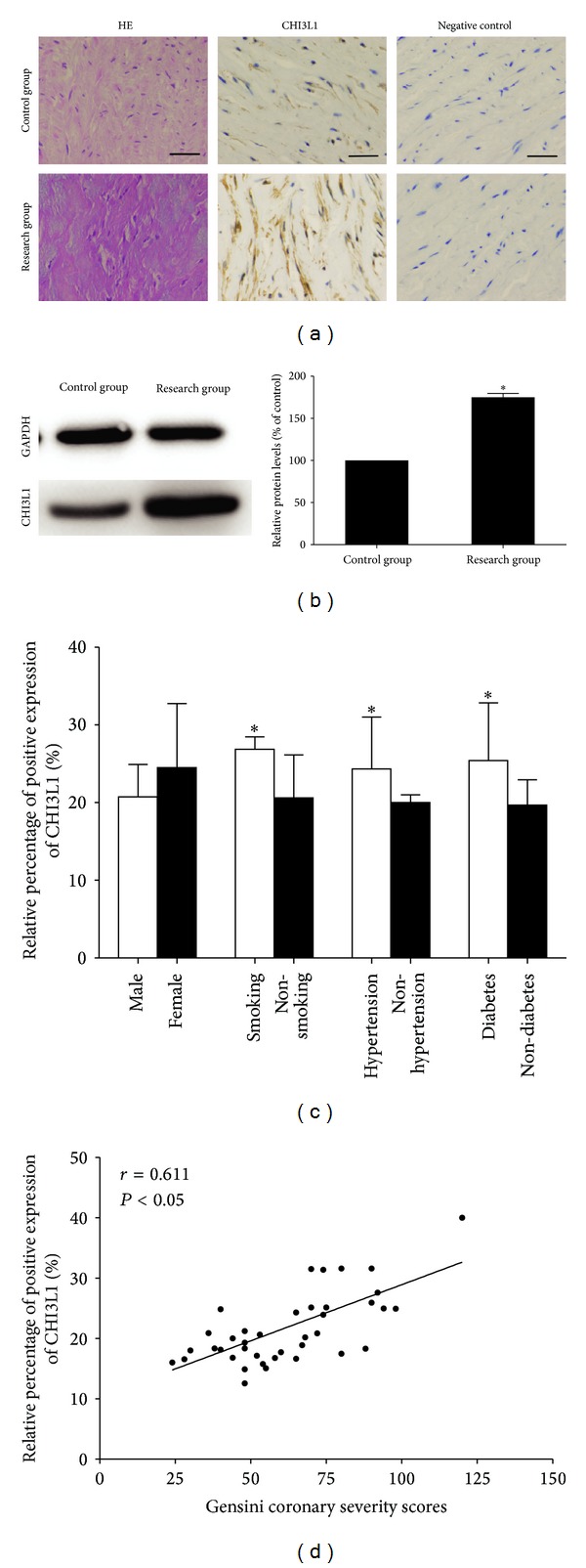
Introduction: The purpose of this study was to investigate the changes of chitinase 3-like 1 (CHI3L1) in the aorta of patients with coronary atherosclerosis and to determine whether inhibition of CHI3L1 by lentivirus-mediated RNA interference could stabilize atherosclerotic plaques in apolipoprotein E-knockout (ApoE(-/-)) mice.

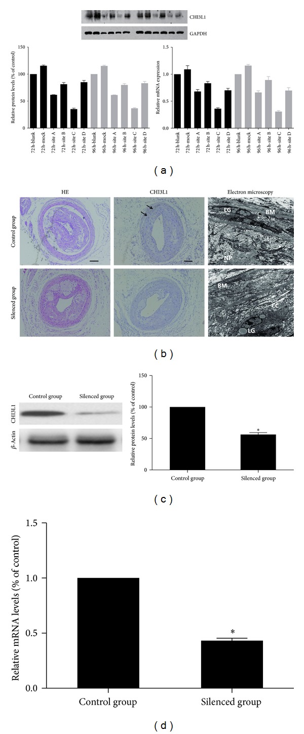
Methods: We collected discarded aortic specimens from patients undergoing coronary artery bypass graft surgery and renal arterial tissues from kidney donors. A lentivirus carrying small interfering RNA targeting the expression of CHI3L1 was constructed. Fifty ApoE(-/-) mice were divided into control group and CHI3L1 gene silenced group. A constrictive collar was placed around carotid artery to induce plaques formation. Then lentivirus was transfected into carotid plaques.

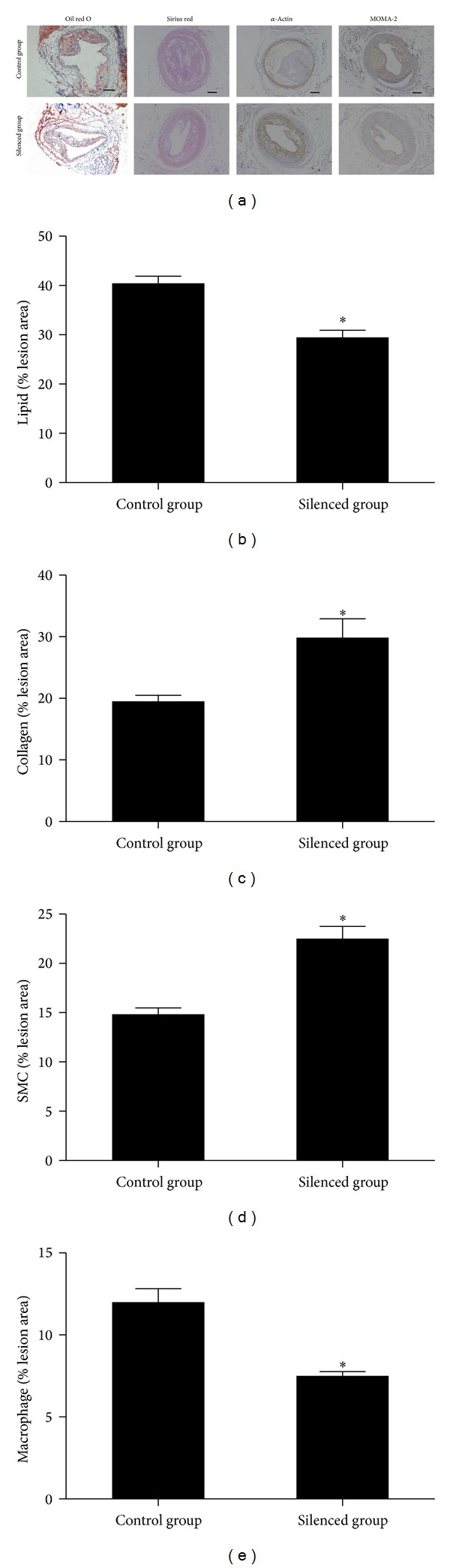
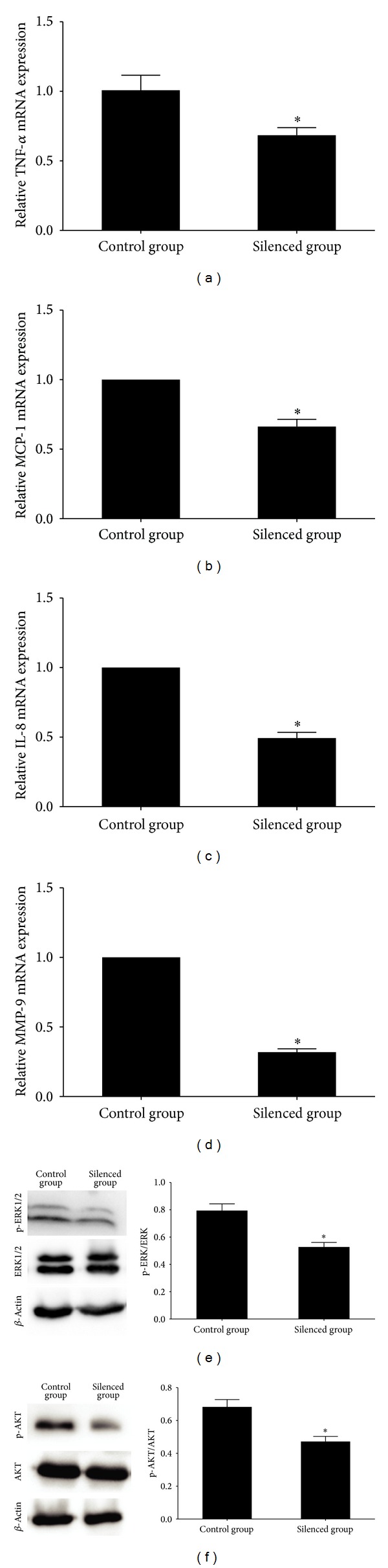
Results: We found that CHI3L1 was overexpressed in aorta of patients with atherosclerosis and its expression was correlated with the atherosclerotic risk factors. After lentivirus transduction, mRNA and protein expression of CHI3L1 were attenuated in carotid plaques, leading to reduced plaque content of lipids and macrophages, and increased plaque content of collagen and smooth muscle cells. Moreover, CHI3L1 gene silencing downregulated the expression of local proinflammatory mediators.

Conclusions: CHI3L1 is overexpressed in aorta from patients with atherosclerosis and the lentivirus-mediated CHI3L1 gene silencing could represent a new strategy to inhibit plaques progression.


Figures

Figure 1
Figure 1
(a) Immunohistochemical staining of CHI3L1 on sections of arterial vessels in control group and research group. The expression of CHI3L1 was increased in the arterial specimens of CAD patients in research group. (scale  bars = 100 μm) (b) Western blot analysis and quantification of CHI3L1 protein expression in control group and research group. The levels of CHI3L1 protein expression were higher in research group than in control group. *P < 0.05 versus control group. (c) Quantitative analysis of arterial CHI3L1 expression in research group patients according to gender, smoking, hypertension, and diabetes mellitus. The expression levels of CHI3L1 were elevated in smokers and patients with hypertension or diabetes mellitus, whereas gender had no significant effect. *P < 0.05. (d) Relationship between arterial CHI3L1 expression and coronary severity scores. The arterial CHI3L1 expression levels were significantly correlated with coronary severity Gensini scores. Each point represents one patient.
Figure 2
Figure 2
(a) Target site screening for CHI3L1 by western blot analysis and real-time RT-PCR in RAW264.7 cells. The RAW264.7 cell line was transfected with lentivirus expressing different CHI3L1 siRNAs, and gene silencing analysis showed that site C lentivirus was the most effective vector in blocking CHI3L1 expression. (b) The immunohistochemical staining of CHI3L1 and electron microscopy in control group and silenced group. In the control group CHI3L1 expression (arrow) could be demonstrated according to the immunohistochemical staining. However, little CHI3L1 was expressed in silenced group. For electron microscopy, in control group most of the endothelial cells denudated and there were a large number of lipid granules (LG) under the basement membrane (BM) in the vessel wall. The atherosclerotic plaques were occupied with necrotic particles (NP), calcification crystals (CC), and cellular debrises. However, in silenced group the number of lipid granules was relatively decreased. (scale  bars = 100 μm) (c) Western blot analysis and quantification of CHI3L1 protein expression in control group and silenced group. The levels of CHI3L1 protein expression were higher in control group than in silenced group. (d) Real-time RT-PCR quantification of CHI3L1 mRNA expression in control group and silenced group. *P < 0.05 versus control group.
Figure 3
Figure 3
CHI3L1 gene silencing influenced plaque composition and stability. (a) Cross-sections of mice carotid arteries in the control group and silenced group were stained for lipids (oil red O), collagen (Sirius red), SMCs (α-actin), and macrophages (MOMA-2). The relative content of lipids (b), collagen (c), SMCs (d), and macrophages (e) in the plaque tissues. The relative contents of lipids and macrophages in plaque tissues were significantly lower in silenced group than in control group. However, the relative collagen and SMCs contents in plaque tissues were increased in silenced group than in control group. *P < 0.05 versus control group.
Figure 4
Figure 4
(a)–(d) Real-time RT-PCR quantification of inflammatory cytokines mRNA expression in mice carotid plaques in control group and silenced group. The silenced group showed lower mRNA expression levels of TNF-α, MCP-1, IL-8, and MMP-9, compared with the control group, and inhibition of CHI3L1 reduced the inductions of proinflammatory cytokines. (e)-(F) Western blot analysis and quantification of ERK1/2, phospho-ERK1/2, AKT, and phospho-AKT protein expression in control group and silenced group. The protein expression levels of phospho-ERK1/2 and phospho-AKT were decreased in silenced group compared with that in control group. *P < 0.05 versus control group.

References

    1. Ross R. Atherosclerosis—an inflammatory disease. The New England Journal of Medicine. 1999;340(2):115–126. - PubMed
    1. Nakashima Y, Raines EW, Plump AS, Breslow JL, Ross R. Upregulation of VCAM-1 and ICAM-1 at atherosclerosis-prone sites on the endothelium in the apoE-deficient mouse. Arteriosclerosis, Thrombosis, and Vascular Biology. 1998;18(5):842–851. - PubMed
    1. Rathcke CN, Vestergaard H. YKL-40—an emerging biomarker in cardiovascular disease and diabetes. Cardiovascular Diabetology. 2009;8, article 61 - PMC - PubMed
    1. Lee CG, Da Silva CA, Dela Cruz CS, et al. Role of chitin and chitinase/chitinase-like proteins in inflammation, tissue remodeling, and injury. Annual Review of Physiology. 2011;73:479–501. - PMC - PubMed
    1. Chupp GL, Lee CG, Jarjour N, et al. A chitinase-like protein in the lung and circulation of patients with severe asthma. The New England Journal of Medicine. 2007;357(20):2016–2027. - PubMed

Publication types

MeSH terms