

Characteristics of intraretinal deposits in acute central serous chorioretinopathy
- PMID: 24729682
- PMCID: PMC3979800
- DOI: 10.2147/OPTH.S48894
Characteristics of intraretinal deposits in acute central serous chorioretinopathy
Abstract
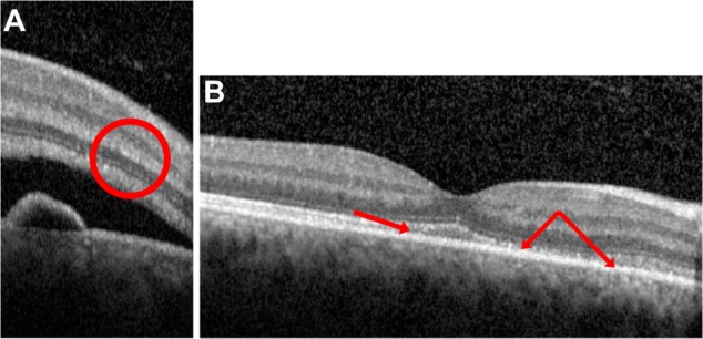
Purpose: To describe the temporal and spatial characteristics of intraretinal deposits in patients with acute central serous chorioretinopathy (CSC) using spectral domain optical coherence tomography (OCT).
Materials and methods: We retrospectively reviewed the medical records of all patients that presented with acute CSC to Weill Cornell Medical College from January 2012 to May 2013. Acute CSC was defined as a diagnosis of CSC within 4 months of the onset of symptoms. Only one eye per patient was included in the study. Each patient was imaged with spectral domain OCT at the initial office visit. The decision to reimage these patients was made by the treating physician.
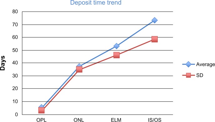
Results: A total of 25 patients (25 eyes; 17 men and eight nonpregnant women) were included in this review. Seven of 25 patients (28%) demonstrated intraretinal deposits within the outer plexiform layer during the initial OCT, with deposits appearing as early as the same day as the onset of symptoms. A total of 25 of 25 patients (100%) demonstrated intraretinal deposits in the outer nuclear layer upon initial (76%) or follow-up OCT, as early as 2 days after the onset of symptoms. A total of 24 of 25 patients (96%) demonstrated deposits in the external limiting membrane upon a follow-up OCT, as early as 7 days from symptoms appearing. A total of 24 of 25 patients (96%) developed intraretinal deposits in the inner segment/outer segment layer upon follow-up OCT, appearing as early as 14 days after symptom onset. At the time of resolution of subretinal fluid, 20 of 25 patients (80%) demonstrated intraretinal deposits.
Conclusion: Intraretinal deposits are present in the outer retinal layers in patients with acute CSC, with the deposits appearing progressively deeper within the retina as the condition evolves. Upon resolution of subretinal fluid, the deposits slowly resolve.
Keywords: acute central serous chorioretinopathy; intraretinal deposits; spectral domain OCT.
Figures

References
-
- Wang M, Munch IC, Hasler PW, Prünte C, Larsen M. Central serous chorioretinopathy. Acta Ophthalmol. 2008;86(2):126–145. - PubMed
-
- Spaide RF, Campeas L, Haas A, et al. Central serous chorioretinopathy in younger and older adults. Ophthalmology. 1996;103(12):2070–2079. - PubMed
-
- Iida T, Hagimura N, Sato T, Kishi S. Evaluation of central serous chorioretinopathy with optical coherence tomography. Am J Ophthalmol. 2000;129(1):16–20. - PubMed
Publication types
LinkOut - more resources
Full Text Sources
Other Literature Sources
