

Review
doi: 10.2147/CCIDE.S41621.
eCollection 2014.
Clinical utility of dental cone-beam computed tomography: current perspectives
Affiliations
- PMID: 24729729
- PMCID: PMC3979889
- DOI: 10.2147/CCIDE.S41621
Item in Clipboard
Review
Clinical utility of dental cone-beam computed tomography: current perspectives
Clin Cosmet Investig Dent.
.
Abstract
Panoramic radiography and computed tomography were the pillars of maxillofacial diagnosis. With the advent of cone-beam computed tomography, dental practice has seen a paradigm shift. This review article highlights the potential applications of cone-beam computed tomography in the fields of dental implantology and forensic dentistry, and its limitations in maxillofacial diagnosis.
Keywords: computed tomography; cone-beam computed tomography; dental implants; panoramic radiography.
Figures
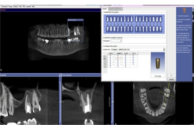
Virtual implant planning by selecting the desired implant from an implant library.
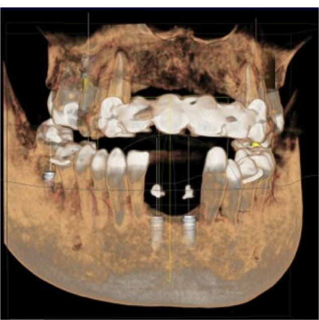
Gutta percha used as a radiographic template.
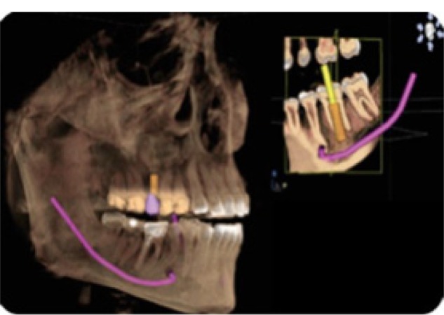
Color coding of the mandibular nerve on Sirona software (Sirona Dental GmbH, Salzburg, Austria).
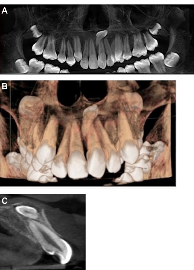
CBCT images (panoramic/3D reconstruction/cross-sectional) eases treatment planning for impacted mesiodens. Notes: (A) Panoramic radiograph showing mesiodens. (B) 3D reconstruction showing impacted mesiodens. (C) Cross sectional view showing relation of impacted mesiodens with central incisor.
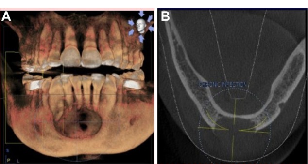
Large radiolucent lesion in the anterior mandible with perforation of the labial cortical plates, as seen on CBCT. Notes: (A) 3D reconstruction showing extensive radiolucent lesion in anterior mandible. (B) Loss of buccal cortical plate clearly demonstrated on axial view. Abbreviation: CBCT, cone-beam computed tomography.
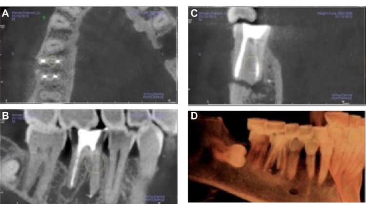
CBCT image showing the missed canal in the mandibular molar. Notes: Images courtesy of Dr Niranjan Vatkar, endodontist, Pune, India. (A) Missed canal seen on axial view. (B) Sagittal view showing large radiolucent lesion with mandibular first molar. (C) Cross sectional view showing missed canal. (D) 3D reconstruction showing osteolytic lesion with mandibular first molar. Abbreviation: CBCT, cone-beam computed tomography.
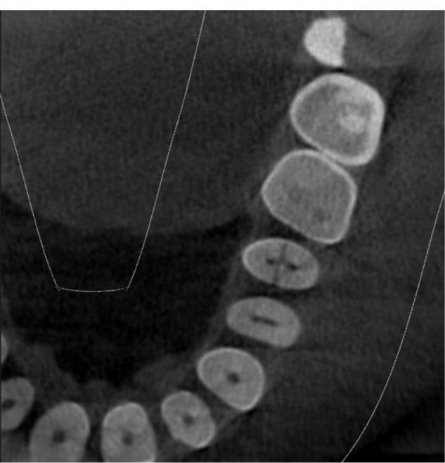
Axial view of CBCT showing the fracture line on the upper-right second premolar. Abbreviation: CBCT, cone-beam computed tomography.
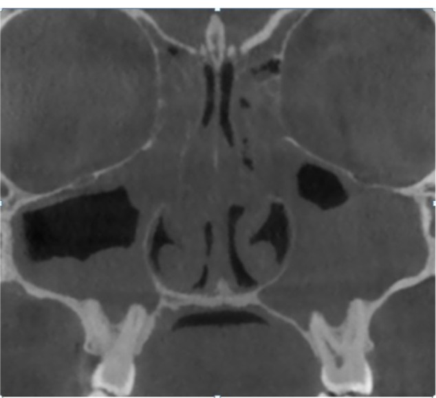
CBCT coronal image showing pansinusitis. Note: Image courtesy of Dr Shikha Rathi Diplomat (AAOMR, USA). Abbreviations: CBCT, cone-beam computed tomography; AAOMR, American Academy of Oral and Maxillofacial Radiology.
References
-
- Tyndall DA, Rathore S. Cone-beam CT diagnostic applications: caries, periodontal bone assessment, and endodontic applications. Dent Clin North Am. 2008;52(4):825–841. vii. - PubMed
-
- Zoller JE, Neugebauer J. Cone-Beam Volumetric Imaging in Dental, Oral and Maxillofacial Medicine: Fundamentals, Diagnostics and Treatment Planning. Chicago, IL: Quintessence Publishing; 2008.
-
- Tetradis S, Anstey P, Graff-Radford S. Cone beam computed tomography in the diagnosis of dental disease. J Calif Dent Assoc. 2010;38(1):27–32. - PubMed
-
- Tyndall DA, Price JB, Tetradis S, Ganz SD, Hildebolt C, Scarfe WC, American Academy of Oral and Maxillofacial Radiology Position statement of the American Academy of Oral and Maxillofacial Radiology on selection criteria for the use of radiology in dental implantology with emphasis on cone beam computed tomography. Oral Surg Oral Med Oral Pathol Oral Radiol. 2012;113(6):817–826. - PubMed
-
- Macleod I, Heath N. Cone-beam computed tomography (CBCT) in dental practice. Dent Update. 2008;35(9):590–592. - PubMed
Publication types
LinkOut - more resources
Full Text Sources
Other Literature Sources
