

Hypoxia-induced endothelial damage and microthrombosis in myocardial vessels of newborn landrace/large white piglets
- PMID: 24729978
- PMCID: PMC3960513
- DOI: 10.1155/2014/619284
Hypoxia-induced endothelial damage and microthrombosis in myocardial vessels of newborn landrace/large white piglets
Abstract
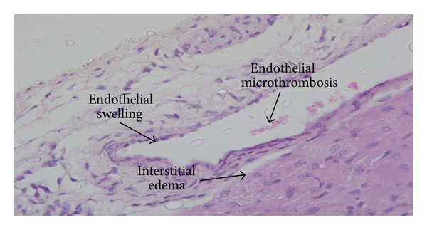
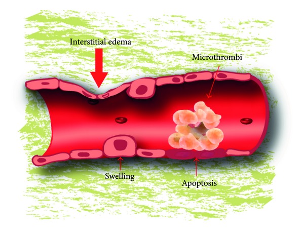
Objective: Evaluating the presence of endothelial changes in myocardial vessels in an experimental model of hypoxia and resuscitation in newborn piglets.
Methods: Fifty male Landrace/Large White neonatal piglets were studied: ten of them were allocated in group A (control group, SHAM-operated). In group B (forty animals, experimental group) normocapnic hypoxia was induced by decreasing inspired concentration of O2 to 6%-8%. When the animals developed bradycardia or severe hypotension, reoxygenation was initiated. The animals of group B were allocated in 4 subgroups of 10, according to the concentration of O2 they were resuscitated with (groups 1, 2, 3, and 4 received 18%, 21%, 40%, and 100% O2, resp.).
Results: Control group animals did not show any significant endothelial lesions. Contrarily, endothelial lesions were detected in all experimental group cases. When these lesions were analyzed in the different heart zones, no significant difference in their incidence was observed; analyzing the frequency in the animals of the 4 subgroups, only microthrombosis showed a higher frequency in animals in groups 4 and 3.
Conclusions: Endothelial damage represents a diffuse pathological feature in the myocardial vessels of piglets subjected to normocapnic hypoxia and resuscitation suggesting a possible role of hyperoxygenation in aggravating endothelial damage.
Figures
References
-
- Sarafidis K. Renal consequences of perinatal asphyxia: the role of urinary biomarkers. In: Fanos V, Chevalier RL, Faa G, Cataldi L, editors. Developmental Nephrology: From Embryology to Metabolomics. Hygeia Press; 2011. pp. 181–193.
-
- Fanos V, Atzori L, Dessì A, D’Aloia E, Finco G, Faa G. The kidney in post-asphytic syndrome: state of the art. In: Fanos V, Chevalier RL, Faa G, Cataldi L, editors. Developmental Nephrology: From Embryology to Metabolomics. Hygeia Press; 2011. pp. 159–179.
-
- Perlman JM, Tack ED, Martin T, Shackelford G, Amon E. Acute systemic organ injury in term infants after asphyxia. The American Journal of Diseases of Children. 1989;143(5):617–620. - PubMed
-
- Beal AL, Cerra FB. Multiple organ failure syndrome in the 1990s: systemic inflammatory response and organ dysfunction. Journal of the American Medical Association. 1994;271(3):226–233. - PubMed
Publication types
MeSH terms
LinkOut - more resources
Full Text Sources
Other Literature Sources
