. 2014 May 13;53(18):3052-61.
doi: 10.1021/bi500252w. Epub 2014 Apr 30.

Structural analysis of replication protein A recruitment of the DNA damage response protein SMARCAL1

Affiliations

Structural analysis of replication protein A recruitment of the DNA damage response protein SMARCAL1

Michael D Feldkamp et al. Biochemistry. .

Abstract

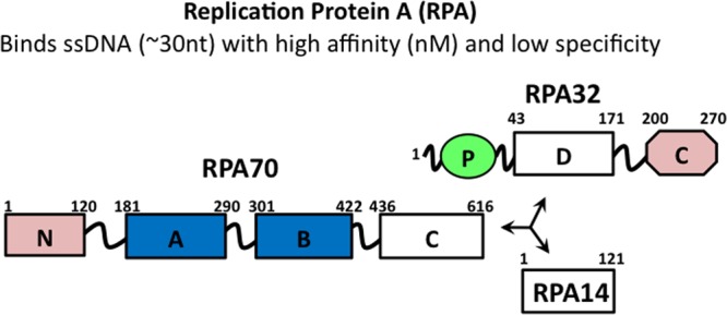
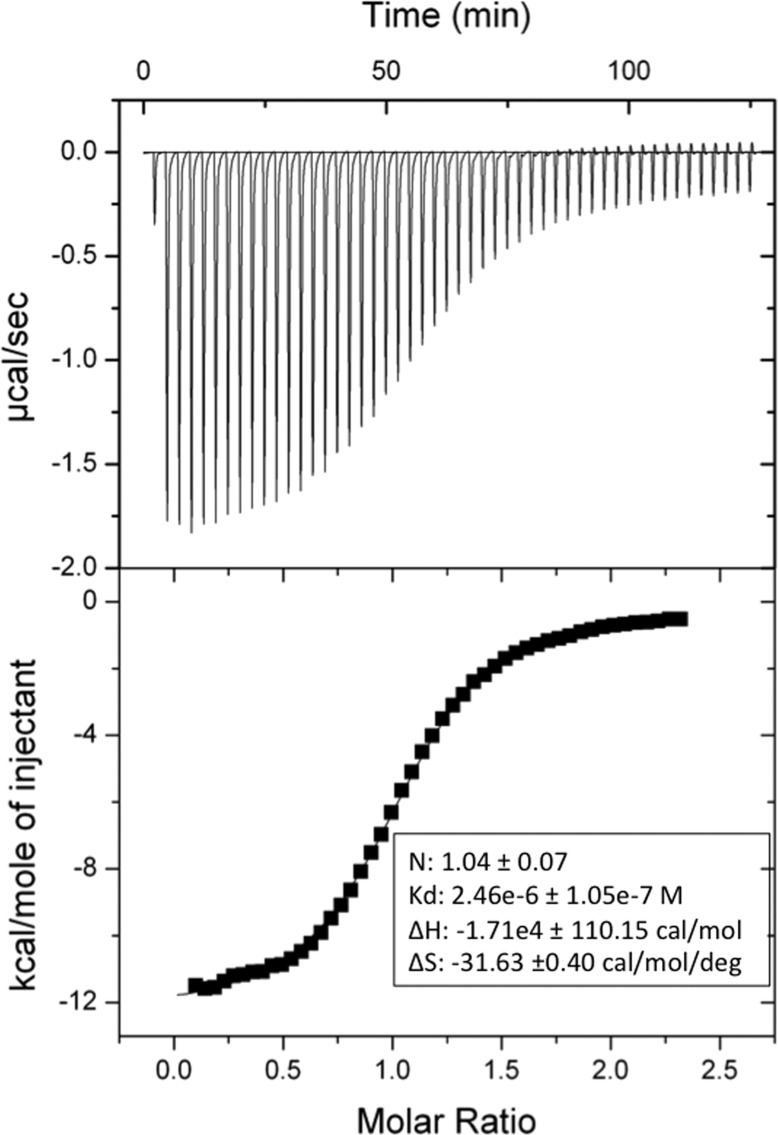
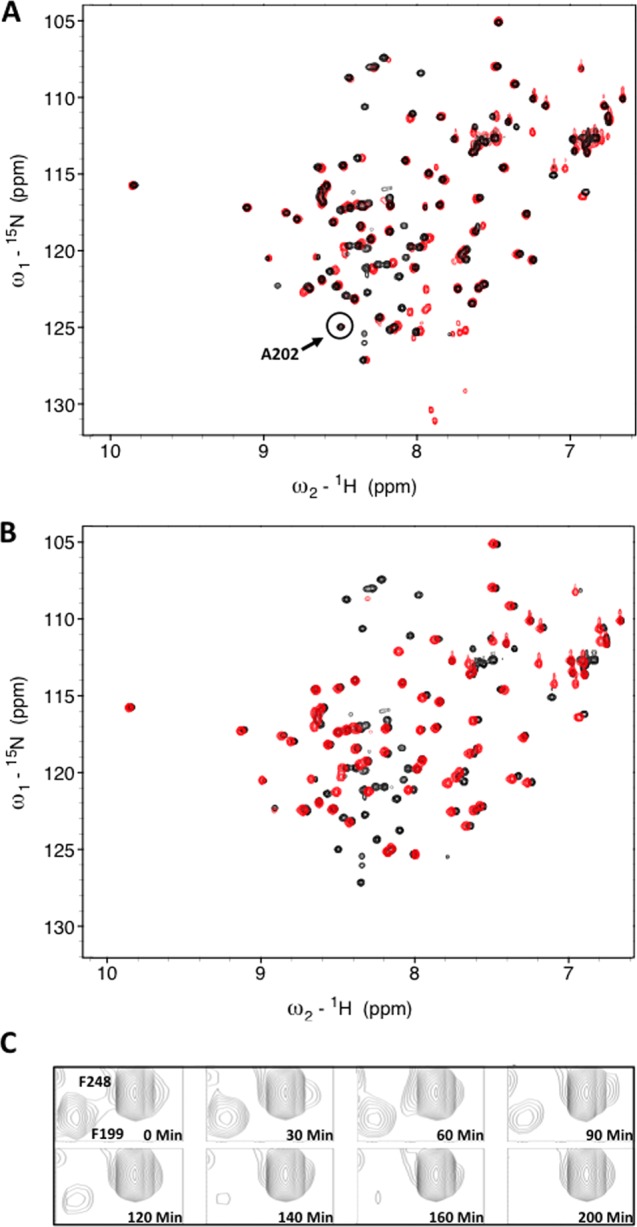
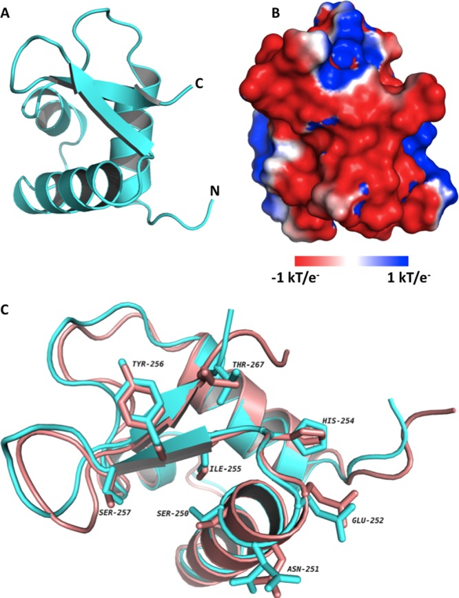
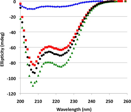
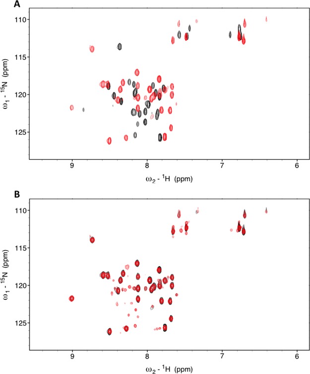
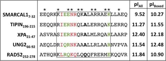
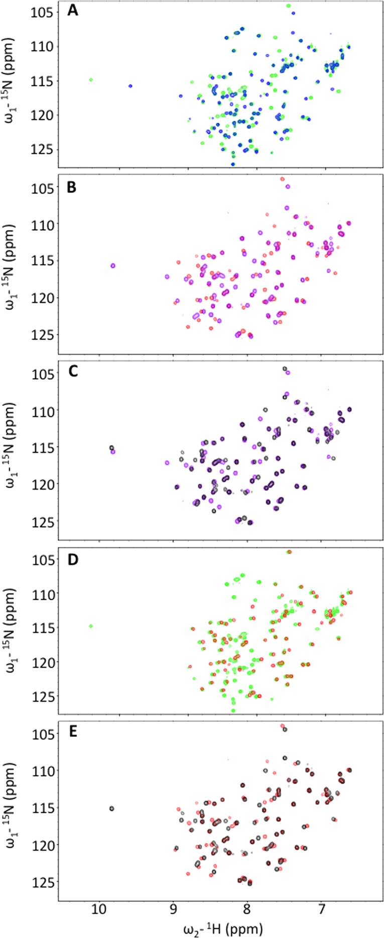
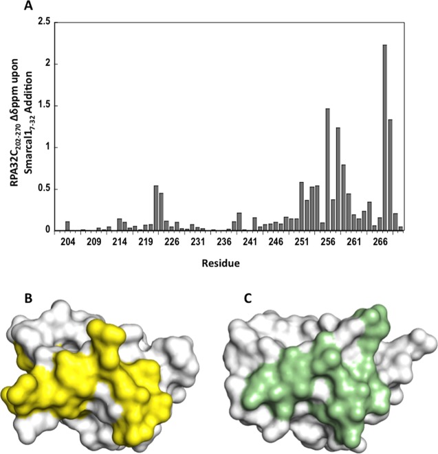
SWI/SNF-related, matrix-associated, actin-dependent regulator of chromatin, subfamily A-like1 (SMARCAL1) is a recently identified DNA damage response protein involved in remodeling stalled replication forks. The eukaryotic single-strand DNA binding protein replication protein A (RPA) recruits SMARCAL1 to stalled forks in vivo and facilitates regression of forks containing leading strand gaps. Both activities are mediated by a direct interaction between an RPA binding motif (RBM) at the N-terminus of SMARCAL1 and the C-terminal winged-helix domain of the RPA 32 kDa subunit (RPA32C). Here we report a biophysical and structural characterization of the SMARCAL1-RPA interaction. Isothermal titration calorimetry and circular dichroism spectroscopy revealed that RPA32C binds SMARCAL1-RBM with a Kd of 2.5 μM and induces a disorder-to-helix transition. The crystal structure of RPA32C was refined to 1.4 Å resolution, and the SMARCAL1-RBM binding site was mapped on the structure on the basis of nuclear magnetic resonance chemical shift perturbations. Conservation of the interaction surface to other RBM-containing proteins allowed construction of a model for the RPA32C/SMARCAL1-RBM complex. The implications of our results are discussed with respect to the recruitment of SMARCAL1 and other DNA damage response and repair proteins to stalled replication forks.


Figures

Figure 1
Figure 1
Subunit and domain structure of RPA. OB-fold domains are depicted as rectangles, and the winged helix–turn–helix domain is depicted as an octagon and the disordered phosphorylation domain as a green oval. The high-affinity ssDNA binding domains are colored blue and the primary protein recruitment domains pink. Trimerization is mediated by RPA70C, -32D, and -14.
Figure 2
Figure 2
Interaction of SMARCAL1 with RPA32C. Isothermal titration calorimetry binding isotherm for titration of SMARCAL1 into RPA32C showing the raw heat release (top) and the integrated heat release (bottom). The experiment was performed at 25 °C with a concentration of 60 μM RPA32172–270 in the cell and 10 μL additions of 800 μM SMARCAL11–32.
Figure 3
Figure 3
(A) 15N–1H HMQC spectra of RPA32C. 15N–1H SOFAST HMQC spectra of RPA32172–270 before (black) and after digestion with (red) Proteinase K. The most N-terminal residue not perturbed by the protease, A202, is highlighted by the circle. (B) Superposition of the 15N–1H SOFAST HMQC spectra of RPA32172–270 (black) and RPA32C (red). (C) A select region of the spectrum with multiple time points to demonstrate residues that are either protected (F248) or digested (F199).
Figure 4
Figure 4
Crystal structure of RPA32C. (A) Ribbon diagram of the X-ray crystal structure of RPA32C. (B) Electrostatic surface potential of RPA32C calculated with APBS. The orientation is identical to that shown in panel A and was selected to show the highly acidic peptide binding site. (C) Superposition of the NMR structure from the RPA32C complex with the UNG2 peptide (salmon) with the crystal structure of free RPA32C (cyan). The orientation is similar to that in panels A and B, with a 45° rotation about the Z axis. A selection of side chains of RPA32C are highlighted, corresponding to those residues in the structure of the complex within 3.5 Å of the UNG2 peptide.
Figure 5
Figure 5
CD spectra of RPA32C, SMARCAL11–32, and the RPA32C–SMARCAL11–32 complex. CD spectra acquired at 25 °C for SMARCAL11–32 alone (blue diamonds), RPA32C alone (red squares), and the SMARCAL11–32–RPA32C complex (green triangles). The sum of the spectra for RPA32C and SMARCAL11–32 is shown with black circles.
Figure 6
Figure 6
NMR analysis of [15N]SMARCAL1 with RPA32C. (A) 15N–1H SOFAST HMQC spectra of [15N]SMARCAL11–32 in the absence (black) and presence (red) of RPA32C. (B) 15N–1H SOFAST HMQC spectra of [15N]SMARCAL11–32 in complex with RPA32C obtained before (black) and after (red) a 50 min Proteinase K digestion.
Figure 7
Figure 7
Sequence alignment of RPA32C target interaction motifs. The asterisks above the SMARCAL1-RMB sequence identify the SMARCAL1-RBM residues in contact with RPA32C in our RosettaDock model. Residues colored green and red represent those that are conserved and highly conserved, respectively. The residues corresponding to the critical alanine residue at position 14 in SMARCAL1 are highlighted in bold. The box is drawn to show the residues that correspond to the RPA32C binding region in the NMR structure of the complex with UNG2. The two columns at right list the pI values of all residues in the motif (pIall) and of only residues in the box (pIbox). The alignment was generated using ClustalW.
Figure 8
Figure 8
NMR analysis to define the minimal regions required for formation of the RPA32C–SMARCAL1 complex. Superposition of pairs of 15N–1H SOFAST HMQC spectra. (A) RPA32172–270 in the absence (blue) and presence of SMARCAL11–32 (green). (B) RPA32C in the absence (purple) and presence of SMARCAL11–32 (red). (C) RPA32C in the absence (purple) and presence of SMARCAL1-RBM (black). (D) RPA32172–270 (green) and RPA32C (red) in the presence of SMARCAL11–32. (E) RPA32C in the presence of SMARCAL11–32 (red) and SMARCAL1-RBM (black).
Figure 9
Figure 9
NMR chemical shift perturbations in RPA32C induced by the binding of SMARCAL1-RBM. (A) Chemical shift perturbations of [15N]RPA32C induced by the binding of the SMARCAL1 peptide. (B) Surface representation of RPA32C with the significant chemical shift perturbations from panel A colored yellow. (C) Surface representation of RPA32C from the UNG2 peptide complex with RPA32C residues within 3.5 Å of the peptide colored green.
Figure 10
Figure 10
Model of the RPA32C–SMARCAL1-RBM complex. The top left panel shows a ribbon representation of the RosettaDock model of RPA32C (cyan) in complex with the SMARCAL1-RMB peptide (yellow). The top right panel shows an overlay of the structure shown in the top left panel with the complex of RPA32C (salmon) in complex with the UNG2 peptide (green). The orientation is shifted by 90° with respect to the top[ left panel. The bottom panel lists contacts between SMARCAL1-RBM residues (yellow) and residues in RPA32C (cyan). Contacts were assigned using Contacts of Structural Units analysis.

References

    1. Zegerman P.; Diffley J. F. (2009) DNA replication as a target of the DNA damage checkpoint. DNA Repair 8, 1077–1088. - PubMed
    1. Bansbach C. E.; Betous R.; Lovejoy C. A.; Glick G. G.; Cortez D. (2009) The annealing helicase SMARCAL1 maintains genome integrity at stalled replication forks. Genes Dev. 23, 2405–2414. - PMC - PubMed
    1. Cimprich K. A.; Cortez D. (2008) ATR: An essential regulator of genome integrity. Nat. Rev. Mol. Cell Biol. 9, 616–627. - PMC - PubMed
    1. Coleman M. A.; Eisen J. A.; Mohrenweiser H. W. (2000) Cloning and characterization of HARP/SMARCAL1: A prokaryotic HepA-related SNF2 helicase protein from human and mouse. Genomics 65, 274–282. - PubMed
    1. Yusufzai T.; Kadonaga J. T. (2008) HARP is an ATP-driven annealing helicase. Science 322, 748–750. - PMC - PubMed

Publication types

LinkOut - more resources