. 2014 Apr 14;5(2):50.
doi: 10.1186/scrt438.

Human adipose tissue-resident monocytes exhibit an endothelial-like phenotype and display angiogenic properties

Human adipose tissue-resident monocytes exhibit an endothelial-like phenotype and display angiogenic properties

Amparo Navarro et al. Stem Cell Res Ther. .

Abstract

Introduction: Adipose tissue has the unique property of expanding throughout adult life, and angiogenesis is required for its growth. However, endothelial progenitor cells contribute minimally to neovascularization. Because myeloid cells have proven to be angiogenic, and monocytes accumulate in expanding adipose tissue, they might contribute to vascularization.

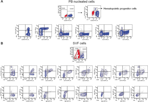
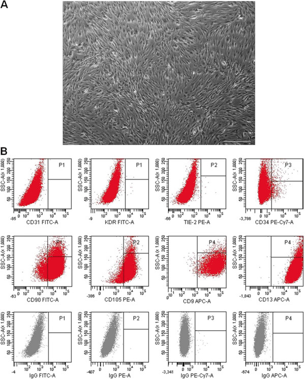
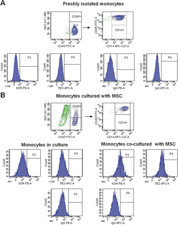
Methods: The stromal vascular fraction (SVF) cells from human adipose tissue were magnetically separated according to CD45 or CD14 expression. Adipose-derived mesenchymal stromal cells (MSCs) were obtained from SVF CD45- cells. CD14+ monocytes were isolated from peripheral blood (PB) mononuclear cells and then cultured with SVF-derived MSCs. Freshly isolated or cultured cells were characterized with flow cytometry; the conditioned media were analyzed for the angiogenic growth factors, angiopoietin-2 (Ang-2), vascular endothelial growth factor (VEGF), basic fibroblast growth factor (bFGF), hepatocyte growth factor (HGF), granulocyte colony-stimulating factor (G-CSF), and granulocyte macrophage colony-stimulating factor (GM-CSF) with Luminex Technology; their angiogenic capacity was determined in an in vivo gelatinous protein mixture (Matrigel) plug angiogenesis assay.

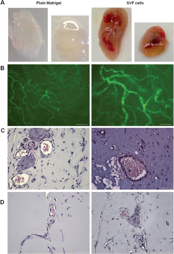
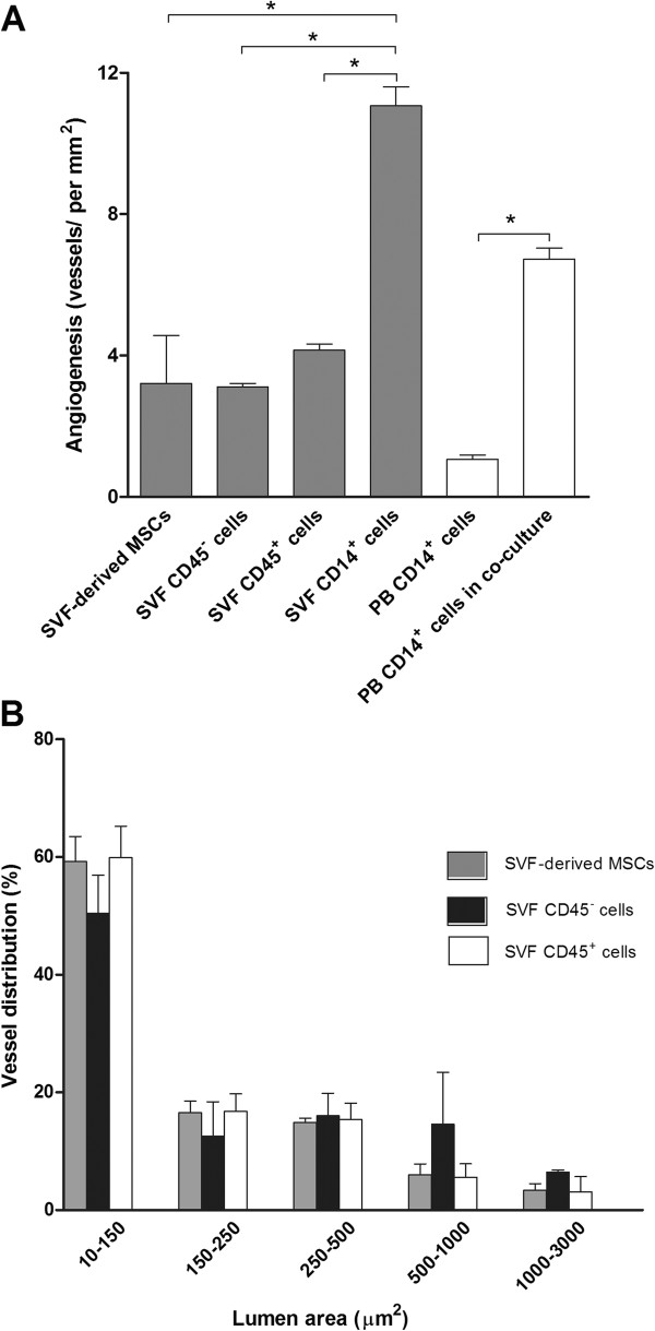
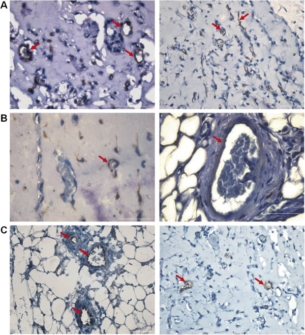
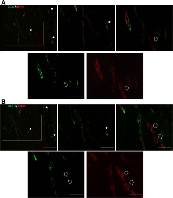
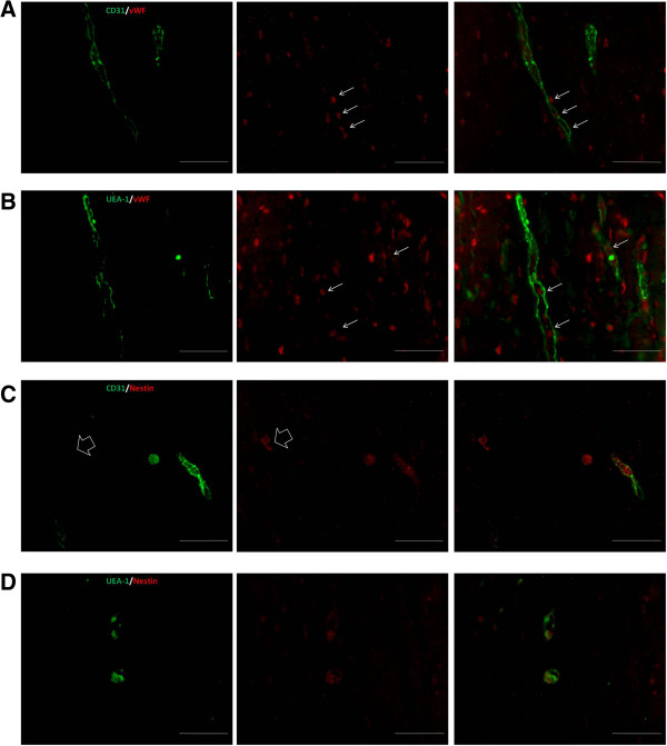
Results: CD45+ hematopoietic cells within the SVF contain CD14+ cells that co-express the CD34 progenitor marker and the endothelial cell antigens VEGF receptor 2 (VEGFR2/KDR), VEGFR1/Flt1, and Tie2. Co-culture experiments showed that SVF-derived MSCs promoted the acquisition of KDR and Tie-2 in PB monocytes. MSCs secreted significant amounts of Ang-2 and HGF, but minimal amounts of bFGF, G-CSF, or GM-CSF, whereas the opposite was observed for SVF CD14+ cells. Additionally, SVF CD14+ cells secreted significantly higher levels of VEGF and bFGF than did MSCs. Culture supernatants of PB monocytes cultured with MSCs contained significantly higher concentrations of VEGF, HGF, G-CSF, and GM-CSF than did the supernatants from cultures without MSCs. Quantitative analysis of angiogenesis at 14 days after implantation demonstrated that neovascularization of the implants containing SVF CD14+ cells or PB monocytes previously co-cultured with MSCs was 3.5 or 2 times higher than that observed in the implants with SVF-derived MSCs. Moreover, immunofluorescence of Matrigel sections revealed that SVF CD14+ cells differentiated into endothelial cells and contributed to vascular endothelium.

Conclusions: The results from this study suggest that adipose tissue-resident monocytes should contribute to tissue vascularization. Because SVF CD14+ cells were more efficient in inducing angiogenesis than SVF-derived MSCs, and differentiated into vascular endothelial cells, they may constitute a new cell source for cell-based therapeutic angiogenesis.


Figures

Figure 1
Figure 1
SVF CD14+ cells exhibit a proangiogenic phenotype. PB nucleated cells (A) were analyzed for CD45 and CD34 expression. Top right, CD34+CD45low cells corresponding to hematopoietic progenitor cells are shown. Bottom, CD45+ cells (blue) were gated and analyzed for CD14 expression and for co-expression of CD14 with CD31, Tie2, KDR, Flt-1, and CD34. The CD14+ cells represented 20% of CD45+ cells, which in turn accounted for 98.5% of the total viable PB nucleated cells. SVF cells (B) were analyzed for CD45 expression, and then the gated CD45+ cells (blue) were analyzed for CD34 and CD14 expression and for the co-expression of CD14 with CD31, CD144, Tie2, KDR, Flt-1, and CD34. Isotype-matched controls are shown. CD45+ cells accounted for 6% of total viable cells. Dot plots of CD14 conjugated with APC or PE correspond to three different samples.
Figure 2
Figure 2
Phenotypic characteristics of adipose-derived MSCs. CD45- SVF cells were isolated by immunomagnetic methods and then cultured in EGM-2 MV to generate MSCs. Morphologic aspect of MSCs at passage 2 (scale bar, 100 μm) is shown in panel (A). Flow-cytometry dot plots demonstrating the expression of a panel of markers in MSCs are shown in (B). Isotype-matched controls are given.
Figure 3
Figure 3
Induction of endothelial markers in circulating CD14+ cells. CD14+ monocytes were isolated from adult PB, cultured in EGM-2 MV with or without MSCs, and then analyzed with flow cytometry. Dot plots from freshly isolated CD14+ cells (A) and after 4 days of co-culture with adipose-derived MSCs (green) (B) are shown. Flow-cytometry histograms in panels A and B show the expression of KDR and Tie2 in gated CD14+ cells. Isotype-matched controls are given.
Figure 4
Figure 4
Angiogenic response induced by SVF cells in the Matrigel-plug assay. Freshly isolated SVF CD45+, CD45-or CD14+ cells, or SVF-derived MSCs were mixed with Matrigel and injected subcutaneously into immunodeficient mice. (A) Macroscopic visualization of Matrigel plugs containing SVF cells or no cells 14 days after implantation. In some experiments, 10 minutes before the Matrigel plugs were harvested, mice were injected into the tail vein with FITC-dextran. Fluorescence microscopy of Matrigel explants allows identifying vessels connected to the circulation (B). Sections from the plugs were stained with H&E to visualize vessel formation (C, D). Scale bars: B, 200 μm (left), 100 μm (right); C and D, 50 μm.
Figure 5
Figure 5
Quantification of neovessels in Matrigel plugs. Freshly isolated SVF cells, SVF-derived MSCs, and PB CD14+ monocytes, freshly isolated or after co-culture with SVF-derived MSCs in a transwell system, were individually embedded in Matrigel at 1 × 106 cells per implant. Matrigel explants were harvested 14 days after implantation and H&E-stained sections were used to enumerate blood vessels (A). The lumen area of newly formed vessels in Matrigel implants containing SVF-derived MSCs or SVF-isolated CD45+ or CD45- cells was determined. Vessel distribution indicates the percentage of vessels according to their lumen area, given as range of values (B). Results are expressed as the mean and SD of five Matrigel implants per test group. MSCs derived from three independent SVFs were used. *P < 0.0001 for differences between groups linked by the brackets.
Figure 6
Figure 6
SVF CD14+ cells incorporate into new vessels. Fourteen days after implantation, Matrigel implants containing SVF CD14+ cells were evaluated for the expression of CD31, CD45, and e-NOS. (A) Representative images for CD31 staining. Arrows indicate the presence of CD31+ endothelial cells in the vessels formed. (B) Human CD45 immunostaining. Note that CD45+ cells are located surrounding blood vessels (left) or adjacent to the endothelium (right). (C) Human e-NOS immunostaining. Arrows indicate positive staining of some blood vessels for e-NOS. Scale bars: A, 50 μm; B, 50 μm (left), 30 μm (right); C, 50 μm.
Figure 7
Figure 7
Matrigel implants contain blood vessels comprising human and mouse endothelial cells. Consecutive sections of Matrigel implants with SVF CD14+ cells were double-immunostained with antibodies against mCD31 (green) and αSMA (red) (A), or Ulex europaeus agglutinin 1 (UEA-1; green) and αSMA (red) (B). The images on the top of the panels A and B show merged images and correspond to the same microscopic field at different magnifications: left, low magnification; middle, medium magnification; right, higher magnification. Images on the bottom (A, B) show green and red fluorescence and correspond to the merged image at the highest magnification. Images in panel B correspond to a consecutive section. The insets in A and B (upper left) show the field at low magnification and asterisks illustrate benchmarks. Note that the green staining associated with murine (A) or human (B) endothelial cells in the adjacent sections is near identical. Double labeling shows a close assembly of αSMA-positive cells to the blood vessels. White arrows indicate αSMA-positive cells lining vascular-like structures which do not express murine (A) or human (B) endothelial cells. Scale bars in A, B (upper left): 200 μm, upper in the middle: 100 μm; upper right and bottom: 50 μm.
Figure 8
Figure 8
Immunofluorescence detection of nestin in Matrigel implants. Representative images of consecutive sections of Matrigel implants with SVF CD14+ cells stained by anti-mouse CD31 (green) and anti-vWF (red) (A), and by UEA-1 (green) and anti-vWF (red) (B). The arrows show colocalization of vWF-positive cells and endothelial cells of murine or human origin. However, in Matrigel implants, other vWF-positive cells not associated with blood vessels are shown. Double-immunofluorescence staining of human nestin (red) and mCD31 (green) (C), and human nestin (red) and UEA-1 (green) (D). In Matrigel implants, nestin was expressed in human and murine endothelial cells, although some nestin-positive cells (arrow) lacking mCD31 were observed. Scale bar, 50 μm.

Similar articles

Cited by

References

    1. Zuk PA, Zhu M, Ashjian P, De Ugarte DA, Huang JI, Mizuno H, Alfonso ZC, Fraser JK, Benhaim P, Hedrick MH. Human adipose tissue is a source of multipotent stem cells. Mol Biol Cell. 2002;13:4279–4295. doi: 10.1091/mbc.E02-02-0105. - DOI - PMC - PubMed
    1. Crandall DL, Haussman GJ, Kral JG. A review of the microcirculation of adipose tissue: anatomic, metabolic, and angiogenic perspectives. Microcirculation. 1997;4:211–232. doi: 10.3109/10739689709146786. - DOI - PubMed
    1. Cao Y. Angiogenesis modulates adipogenesis and obesity. J Clin Invest. 2007;117:2362–2368. doi: 10.1172/JCI32239. - DOI - PMC - PubMed
    1. Shi Q, Rafii S, Wu MH, Wijelath ES, Yu C, Ishida A, Fujita Y, Kothari S, Mohle R, Sauvage LR, Moore MA, Storb RF, Hammond WP. Evidence for circulating bone marrow-derived endothelial cells. Blood. 1998;92:362–367. - PubMed
    1. Bailey AS, Willenbring H, Jiang S, Anderson DA, Schroeder DA, Wong MH, Grompe M, Fleming WH. Myeloid lineage progenitors give rise to vascular endothelium. Proc Natl Acad Sci U S A. 2006;103:13156–13161. doi: 10.1073/pnas.0604203103. - DOI - PMC - PubMed

Publication types