Review
. 2014 May;30(2):153-63. v.
doi: 10.1016/j.hcl.2013.12.005.

Flap reconstruction of the elbow and forearm: a case-based approach

Affiliations
Review

Flap reconstruction of the elbow and forearm: a case-based approach

Joshua M Adkinson et al. Hand Clin. 2014 May.

Abstract

Elbow and forearm wounds have distinct reconstructive requirements, but both require a durable and pliable solution. Pedicle, free fasciocutaneous and muscle, and distant (2-stage) flaps have a role in wound reconstruction in these unique areas. This article presents practical surgical cases as a guide to soft tissue reconstruction of the elbow and forearm.

Keywords: Elbow wounds; Flap reconstruction; Forearm wounds.


Figures

Figure 1
Figure 1
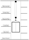
Reconstructive Elevator Adapted from Bennett N, Choudhary S. Why climb a ladder when you can take the elevator? Plast Reconstr Surg. 2000 May;105(6):2266; with permission.
Figure 2
Figure 2
Radial Forearm Pedicle Flap (A) elbow wound after debridement (B) proximally-based radial forearm flap (C) flap inset into wound. (D) post-operative result
Figure 2
Figure 2
Radial Forearm Pedicle Flap (A) elbow wound after debridement (B) proximally-based radial forearm flap (C) flap inset into wound. (D) post-operative result
Figure 2
Figure 2
Radial Forearm Pedicle Flap (A) elbow wound after debridement (B) proximally-based radial forearm flap (C) flap inset into wound. (D) post-operative result
Figure 2
Figure 2
Radial Forearm Pedicle Flap (A) elbow wound after debridement (B) proximally-based radial forearm flap (C) flap inset into wound. (D) post-operative result
Figure 3
Figure 3
Reverse Lateral Arm Pedicle Flap (A) pre-operative image of elbow wound (B) flap markings (C) flap dissection (D) flap inset into wound.
Figure 3
Figure 3
Reverse Lateral Arm Pedicle Flap (A) pre-operative image of elbow wound (B) flap markings (C) flap dissection (D) flap inset into wound.
Figure 3
Figure 3
Reverse Lateral Arm Pedicle Flap (A) pre-operative image of elbow wound (B) flap markings (C) flap dissection (D) flap inset into wound.
Figure 3
Figure 3
Reverse Lateral Arm Pedicle Flap (A) pre-operative image of elbow wound (B) flap markings (C) flap dissection (D) flap inset into wound.
Figure 4
Figure 4
Anterolateral Thigh Free Flap (A) extensive forearm and hand wound after debridement (B) flap markings (C) flap elevation prior to transfer (D) flap inset into wound.
Figure 4
Figure 4
Anterolateral Thigh Free Flap (A) extensive forearm and hand wound after debridement (B) flap markings (C) flap elevation prior to transfer (D) flap inset into wound.
Figure 4
Figure 4
Anterolateral Thigh Free Flap (A) extensive forearm and hand wound after debridement (B) flap markings (C) flap elevation prior to transfer (D) flap inset into wound.
Figure 4
Figure 4
Anterolateral Thigh Free Flap (A) extensive forearm and hand wound after debridement (B) flap markings (C) flap elevation prior to transfer (D) flap inset into wound.
Figure 5
Figure 5
Flexor Carpi Ulnaris Pedicle Flap (A) elbow flexion contracture (B) elbow and forearm wounds after burn contracture release (C) FCU muscle insertion divided and held in forceps (D) FCU muscle reflected proximally for medial epicondyle coverage (E) flap inset and skin graft placed.
Figure 5
Figure 5
Flexor Carpi Ulnaris Pedicle Flap (A) elbow flexion contracture (B) elbow and forearm wounds after burn contracture release (C) FCU muscle insertion divided and held in forceps (D) FCU muscle reflected proximally for medial epicondyle coverage (E) flap inset and skin graft placed.
Figure 5
Figure 5
Flexor Carpi Ulnaris Pedicle Flap (A) elbow flexion contracture (B) elbow and forearm wounds after burn contracture release (C) FCU muscle insertion divided and held in forceps (D) FCU muscle reflected proximally for medial epicondyle coverage (E) flap inset and skin graft placed.
Figure 5
Figure 5
Flexor Carpi Ulnaris Pedicle Flap (A) elbow flexion contracture (B) elbow and forearm wounds after burn contracture release (C) FCU muscle insertion divided and held in forceps (D) FCU muscle reflected proximally for medial epicondyle coverage (E) flap inset and skin graft placed.
Figure 5
Figure 5
Flexor Carpi Ulnaris Pedicle Flap (A) elbow flexion contracture (B) elbow and forearm wounds after burn contracture release (C) FCU muscle insertion divided and held in forceps (D) FCU muscle reflected proximally for medial epicondyle coverage (E) flap inset and skin graft placed.
Figure 6
Figure 6
Brachioradialis Pedicle Flap (A) pre-operative image of elbow wound (B) flap dissection, sparing the dorsal radial sensory nerve (C) flap elevated (D) long-term result after primary closure over the FCU muscle flap.
Figure 6
Figure 6
Brachioradialis Pedicle Flap (A) pre-operative image of elbow wound (B) flap dissection, sparing the dorsal radial sensory nerve (C) flap elevated (D) long-term result after primary closure over the FCU muscle flap.
Figure 6
Figure 6
Brachioradialis Pedicle Flap (A) pre-operative image of elbow wound (B) flap dissection, sparing the dorsal radial sensory nerve (C) flap elevated (D) long-term result after primary closure over the FCU muscle flap.
Figure 6
Figure 6
Brachioradialis Pedicle Flap (A) pre-operative image of elbow wound (B) flap dissection, sparing the dorsal radial sensory nerve (C) flap elevated (D) long-term result after primary closure over the FCU muscle flap.
Figure 7
Figure 7
Latissimus Dorsi Free Flap (A) forearm wound after debridement (B) arterial anatomy of flap (C) free latissimus dorsi muscle flap after inset (D) long-term result.
Figure 7
Figure 7
Latissimus Dorsi Free Flap (A) forearm wound after debridement (B) arterial anatomy of flap (C) free latissimus dorsi muscle flap after inset (D) long-term result.
Figure 7
Figure 7
Latissimus Dorsi Free Flap (A) forearm wound after debridement (B) arterial anatomy of flap (C) free latissimus dorsi muscle flap after inset (D) long-term result.
Figure 7
Figure 7
Latissimus Dorsi Free Flap (A) forearm wound after debridement (B) arterial anatomy of flap (C) free latissimus dorsi muscle flap after inset (D) long-term result.
Figure 8
Figure 8
Intercostal Artery Perforator Pedicle Flap (A) elbow flexion and forearm scar contractures (B) elbow and forearm wound after debridement and adjacent z-plasty (C) flap design and elevation (D) flap inset (E) long-term result.
Figure 8
Figure 8
Intercostal Artery Perforator Pedicle Flap (A) elbow flexion and forearm scar contractures (B) elbow and forearm wound after debridement and adjacent z-plasty (C) flap design and elevation (D) flap inset (E) long-term result.
Figure 8
Figure 8
Intercostal Artery Perforator Pedicle Flap (A) elbow flexion and forearm scar contractures (B) elbow and forearm wound after debridement and adjacent z-plasty (C) flap design and elevation (D) flap inset (E) long-term result.
Figure 8
Figure 8
Intercostal Artery Perforator Pedicle Flap (A) elbow flexion and forearm scar contractures (B) elbow and forearm wound after debridement and adjacent z-plasty (C) flap design and elevation (D) flap inset (E) long-term result.
Figure 8
Figure 8
Intercostal Artery Perforator Pedicle Flap (A) elbow flexion and forearm scar contractures (B) elbow and forearm wound after debridement and adjacent z-plasty (C) flap design and elevation (D) flap inset (E) long-term result.

Similar articles

Cited by

References

    1. Giele H. Soft tissue coverage around the elbow. In: Stanley D, Trail I, editors. Operative Elbow Surgery: Expert Consult. Churchill Livingstone; London, England: 2012. p. 719.
    1. Sharpe F, Stevanovic M, Itamura JM. Soft tissue coverage of the elbow. In: Mirzayan R, Itamura JM, editors. Shoulder and Elbow Trauma. Thieme; New York, NY: 2004. p. 89.
    1. Stevanovic M, Sharpe F. Soft-tissue coverage of the elbow. Plast Reconstr Surg. 2013 Sep;132(3):387e–402e. - PubMed
    1. Jensen M, Moran SL. Soft tissue coverage of the elbow: a reconstructive algorithm. Orthop Clin North Am. 2008 Apr;39(2):251–64. vii. - PubMed
    1. Levin LS. The reconstructive ladder. An orthoplastic approach. Orthop Clin North Am. 1993 Jul;24:393–409. - PubMed

Publication types