. 2014 Apr 14;9(4):e94476.
doi: 10.1371/journal.pone.0094476. eCollection 2014.

Expression profiling of primary and metastatic ovarian tumors reveals differences indicative of aggressive disease

Affiliations

Expression profiling of primary and metastatic ovarian tumors reveals differences indicative of aggressive disease

Alexander S Brodsky et al. PLoS One. .

Abstract

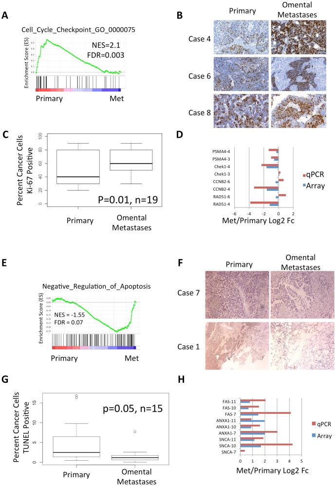
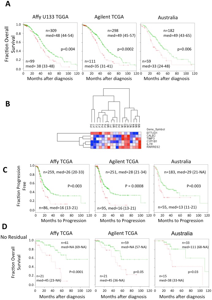
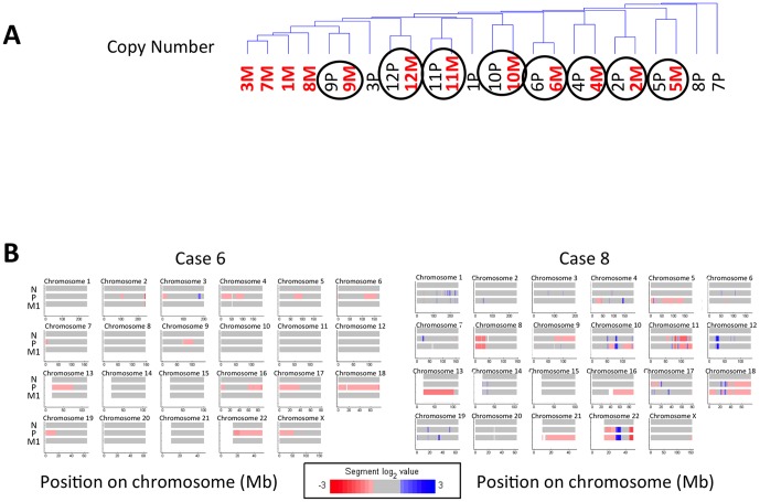
The behavior and genetics of serous epithelial ovarian cancer (EOC) metastasis, the form of the disease lethal to patients, is poorly understood. The unique properties of metastases are critical to understand to improve treatments of the disease that remains in patients after debulking surgery. We sought to identify the genetic and phenotypic landscape of metastatic progression of EOC to understand how metastases compare to primary tumors. DNA copy number and mRNA expression differences between matched primary human tumors and omental metastases, collected at the same time during debulking surgery before chemotherapy, were measured using microarrays. qPCR and immunohistochemistry validated findings. Pathway analysis of mRNA expression revealed metastatic cancer cells are more proliferative and less apoptotic than primary tumors, perhaps explaining the aggressive nature of these lesions. Most cases had copy number aberrations (CNAs) that differed between primary and metastatic tumors, but we did not detect CNAs that are recurrent across cases. A six gene expression signature distinguishes primary from metastatic tumors and predicts overall survival in independent datasets. The genetic differences between primary and metastatic tumors, yet common expression changes, suggest that the major clone in metastases is not the same as in primary tumors, but the cancer cells adapt to the omentum similarly. Together, these data highlight how ovarian tumors develop into a distinct, more aggressive metastatic state that should be considered for therapy development.


Conflict of interest statement

Competing Interests: The authors have declared that no competing interests exist.

Figures

Figure 1
Figure 1. Metastases are more proliferative and include less apoptotic cells than matched primary tumors.
A. GSEA enrichment plot suggests Reactome G2/M cell cycle checkpoints including Chek1, CCNB2, and BUB1 are expressed higher in primary tumors. B. Representative immunohistochemical staining of Ki-67 from three cases suggest higher Ki-67 staining in omental metastases. C. Box plot of the percent cells with positive Ki-67 staining. Paired t-test suggests significant differences between omental metastases and primary tumors from 19 cases. D. qPCR validation of cell cycle checkpoints. Note that genes with significant >1.8 fold changes in the array validate in those same tumors by qPCR. Genes with small expression changes measured by either the array or qPCR may be noisy in those tumors. E. GSEA enrichment plot suggests that negative regulators of apoptosis are up-regulated in metastases. F. TUNEL staining of two representative cases shows increased TUNEL signal in metastases. G. Box plot of the percent of cancer cells as determined by H&E staining with positive TUNEL staining. Paired t-test suggests significance.
Figure 2
Figure 2. Six gene metastasis signature distinguishes aggressive disease in patients and primary and metastatic tumors.
A. Red dotted lines indicate high risk group and the green line indicates the low risk group. For each patient’s tumor, a point was given for each metastasis gene for which higher than median expression was associated with longer survival, and vice versa. Tumors with more than three points were classified as high risk. Only patients who were treated with platinum or taxane chemotherapy were included. Two microarray platforms from TCGA were evaluated (Affymetrix U133A and the Agilent Custom 244K). A second independent dataset on Affymetrix U133 Plus 2 microarrays from the Australian Oncology group was also evaluated , . The median survival is shown with 95% confidence levels in parentheses. B. Hierarchical clustering of the six gene metastasis signature separates the primary and metastatic tumors. Red indicates higher and blue lower expression. C. The six gene metastatic signature distinguishes more aggressive disease using time to progression as a metric. D. The six gene signature predicts survival for cases with no residual disease. Cases in each dataset with no macro disease after surgery were gathered and evaluated for overall survival.
Figure 3
Figure 3. Hierarchical clustering of copy number aberrations and mRNA expression.
A. Most patients cluster by their copy number aberrations (CNAs). The circles indicate matched pairs of primary and metastatic tumors that cluster together with similar patterns of CNAs. B. Representative heat maps of the copy number data from two cases. Many CNAs are found in both primary and metastatic tumors.

References

    1. Lengyel E (2010) Ovarian cancer development and metastasis. Am J Pathol 177: 1053–1064 10.2353/ajpath.2010.100105 - DOI - PMC - PubMed
    1. Bos PD, Zhang XH, Nadal C, Shu W, Gomis RR, et al. (2009) Genes that mediate breast cancer metastasis to the brain. Nature 459: 1005–1009 10.1038/nature08021 - DOI - PMC - PubMed
    1. Paris PL, Hofer MD, Albo G, Kuefer R, Gschwend JE, et al. (2006) Genomic profiling of hormone-naive lymph node metastases in patients with prostate cancer. Neoplasia 8: 1083–1089 10.1593/neo.06421 - DOI - PMC - PubMed
    1. Adib TR, Henderson S, Perrett C, Hewitt D, Bourmpoulia D, et al. (2004) Predicting biomarkers for ovarian cancer using gene-expression microarrays. Br J Cancer 90: 686–692 10.1038/sj.bjc.6601603 - DOI - PMC - PubMed
    1. Hibbs K, Skubitz KM, Pambuccian SE, Casey RC, Burleson KM, et al. (2004) Differential gene expression in ovarian carcinoma: identification of potential biomarkers. Am J Pathol 165: 397–414 10.1016/S0002-9440(10)63306-8 - DOI - PMC - PubMed

Publication types