. 2014 Apr 14;9(4):e94820.
doi: 10.1371/journal.pone.0094820. eCollection 2014.

High resolution systematic digital histological quantification of cardiac fibrosis and adipose tissue in phospholamban p.Arg14del mutation associated cardiomyopathy

Affiliations

High resolution systematic digital histological quantification of cardiac fibrosis and adipose tissue in phospholamban p.Arg14del mutation associated cardiomyopathy

Johannes M I H Gho et al. PLoS One. .

Abstract

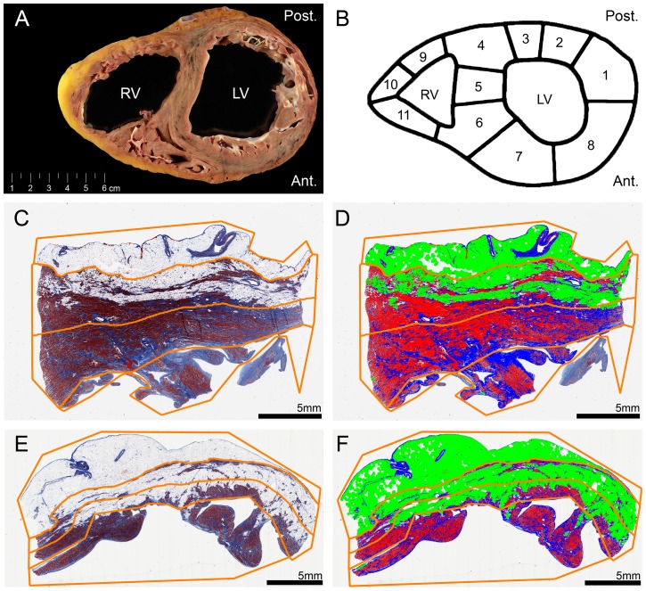
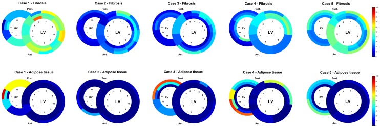
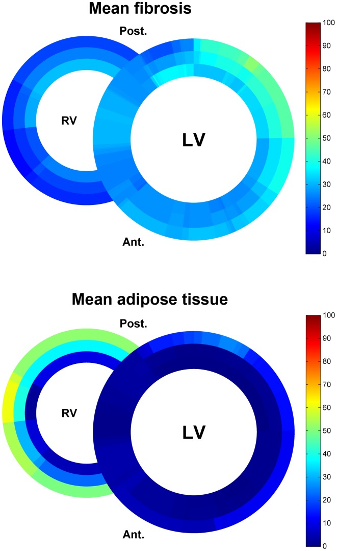
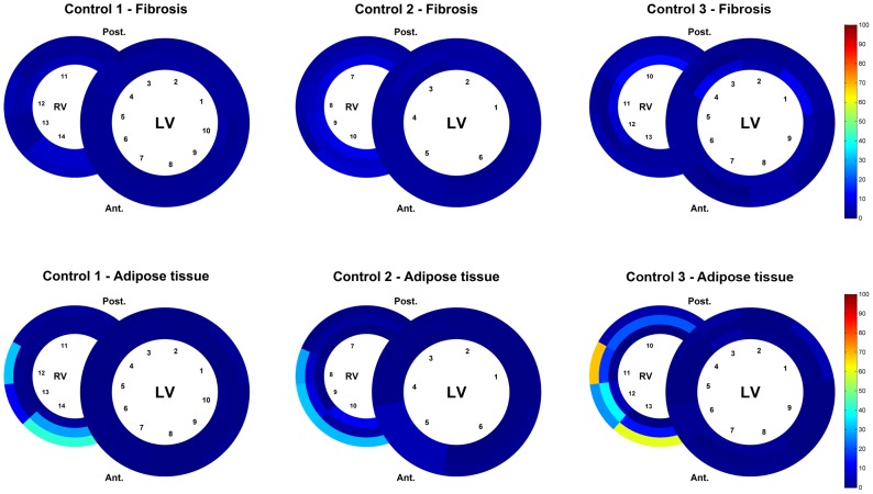
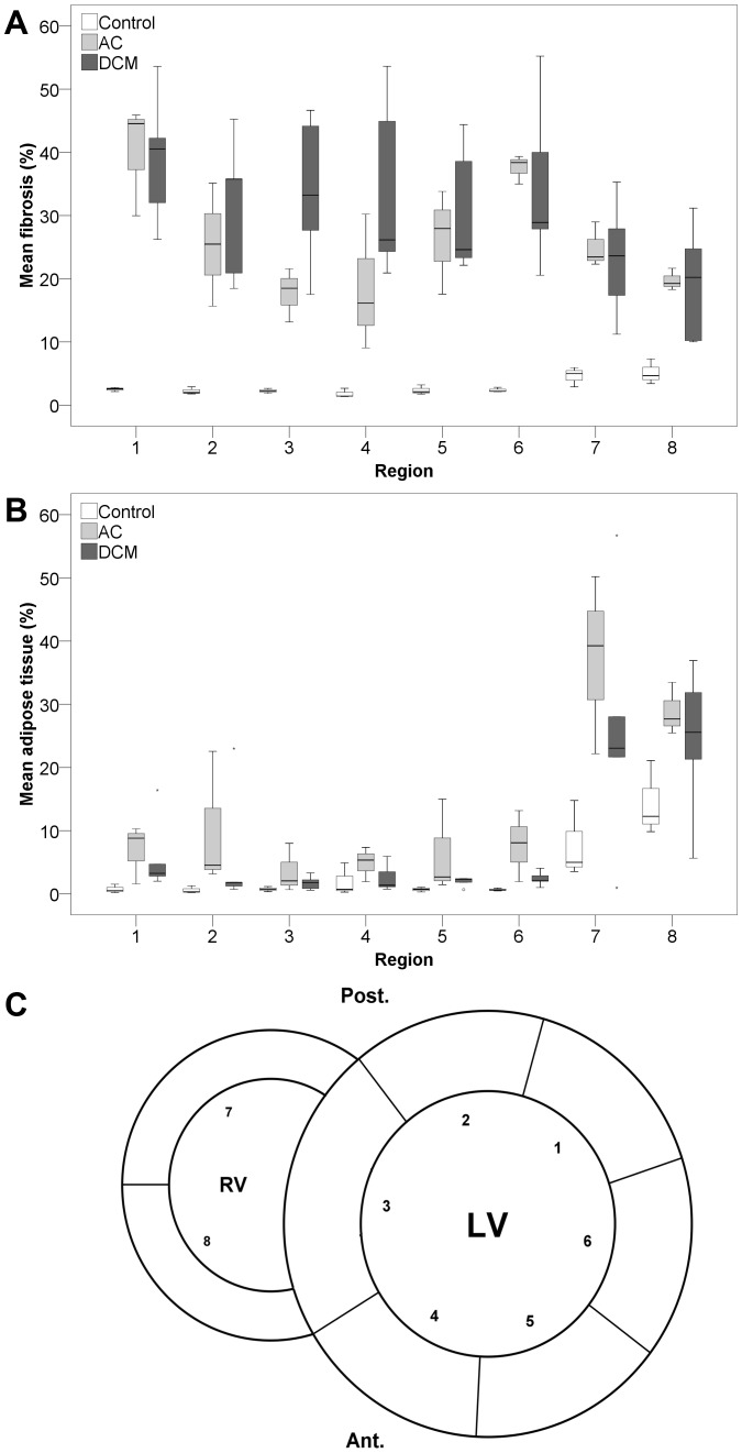
Myocardial fibrosis can lead to heart failure and act as a substrate for cardiac arrhythmias. In dilated cardiomyopathy diffuse interstitial reactive fibrosis can be observed, whereas arrhythmogenic cardiomyopathy is characterized by fibrofatty replacement in predominantly the right ventricle. The p.Arg14del mutation in the phospholamban (PLN) gene has been associated with dilated cardiomyopathy and recently also with arrhythmogenic cardiomyopathy. Aim of the present study is to determine the exact pattern of fibrosis and fatty replacement in PLN p.Arg14del mutation positive patients, with a novel method for high resolution systematic digital histological quantification of fibrosis and fatty tissue in cardiac tissue. Transversal mid-ventricular slices (n = 8) from whole hearts were collected from patients with the PLN p.Arg14del mutation (age 48±16 years; 4 (50%) male). An in-house developed open source MATLAB script was used for digital analysis of Masson's trichrome stained slides (http://sourceforge.net/projects/fibroquant/). Slides were divided into trabecular, inner and outer compact myocardium. Per region the percentage of connective tissue, cardiomyocytes and fatty tissue was quantified. In PLN p.Arg14del mutation associated cardiomyopathy, myocardial fibrosis is predominantly present in the left posterolateral wall and to a lesser extent in the right ventricular wall, whereas fatty changes are more pronounced in the right ventricular wall. No difference in distribution pattern of fibrosis and adipocytes was observed between patients with a clinical predominantly dilated and arrhythmogenic cardiomyopathy phenotype. In the future, this novel method for quantifying fibrosis and fatty tissue can be used to assess cardiac fibrosis and fatty tissue in animal models and a broad range of human cardiomyopathies.


Conflict of interest statement

Competing Interests: The authors have declared that no competing interests exist.

Figures

Figure 1
Figure 1. Overview of methodology.
LV, left ventricle; RV, right ventricle; Ant., anterior; Post., posterior. A, gross showing a transverse heart slice of arrhythmogenic cardiomyopathy. B, transverse slice dissection scheme. CF, examples of digital slide processing. Regions of interest are shown in orange lines (defining the epicardium, compact myocardium divided by an equidistant midline and trabeculated part). C, slide from the left ventricle posterior wall. D, slide C after digital processing. Red: cardiomyocytes. Blue: connective tissue. Pseudo green: adipose tissue. E, slide from the right ventricle lateral wall. F, slide E after digital processing. Red: cardiomyocytes. Blue: connective tissue. Pseudo green: adipose tissue.
Figure 2
Figure 2. Schematic heart slice overview of patients with a clinical phenotype of dilated cardiomyopathy.
The results of the digital quantification in heart slices in percentage of fibrosis or adipose tissue are shown using a color scale. The epicardial region has been excluded from this overview. LV, left ventricle; RV, right ventricle; Ant., anterior; Post., posterior
Figure 3
Figure 3. Schematic heart slice overview of patients with a clinical phenotype of arrhythmogenic cardiomyopathy.
The results of the digital quantification in heart slices in percentage of fibrosis or adipose tissue are shown using a color scale. The epicardial region has been excluded from this overview. LV, left ventricle; RV, right ventricle; Ant., anterior; Post., posterior
Figure 4
Figure 4. Schematic heart slice overview of patients with the PLN p.Arg14del mutation (dilated and arrhythmogenic cardiomyopathy).
The results of the mean percentage of fibrosis or adipose tissue are shown using a color scale. In total 102 heart slides of 8 heart slices were used. LV, left ventricle; RV, right ventricle; Ant., anterior; Post., posterior
Figure 5
Figure 5. Schematic overview of fibrosis and adipose tissue in heart slices of control hearts.
The results of the digital quantification in percentage of fibrosis or adipose tissue are shown using a color scale. The epicardial region has been excluded from the analysis. LV, left ventricle; RV, right ventricle; Ant., anterior; Post., posterior
Figure 6
Figure 6. Boxplots of mean fibrosis and mean adipose tissue in the different regions.
Boxplots of mean percentage of fibrosis (A) and adipose tissue (B) per condition in 8 regions (C) corrected for surface area. Outliers are represented by small circles or stars. AC, arrhythmogenic cardiomyopathy; DCM, dilated cardiomyopathy; LV, left ventricle; RV, right ventricle; Ant., anterior; Post., posterior; 1 =  LV posterolateral wall; 2  =  LV posterior wall; 3  =  interventricular septum; 4  =  LV anterior wall; 5  =  LV anterolateral wall; 6  =  LV lateral wall; 7  =  RV posterior wall; 8  =  RV anterior wall.

Similar articles

Cited by

References

    1. Weber KT, Sun Y, Bhattacharya SK, Ahokas RA, Gerling IC (2013) Myofibroblast-mediated mechanisms of pathological remodelling of the heart. Nature reviews Cardiology 10: 15–26. - PubMed
    1. Marcus FI, McKenna WJ, Sherrill D, Basso C, Bauce B, et al. (2010) Diagnosis of arrhythmogenic right ventricular cardiomyopathy/dysplasia: proposed modification of the task force criteria. Circulation 121: 1533–1541. - PMC - PubMed
    1. Mewton N, Liu CY, Croisille P, Bluemke D, Lima JA (2011) Assessment of myocardial fibrosis with cardiovascular magnetic resonance. Journal of the American College of Cardiology 57: 891–903. - PMC - PubMed
    1. de Jong S, van Veen TA, de Bakker JM, van Rijen HV (2012) Monitoring cardiac fibrosis: a technical challenge. Netherlands heart journal: monthly journal of the Netherlands Society of Cardiology and the Netherlands Heart Foundation 20: 44–48. - PMC - PubMed
    1. Schmitt JP, Kamisago M, Asahi M, Li GH, Ahmad F, et al. (2003) Dilated cardiomyopathy and heart failure caused by a mutation in phospholamban. Science 299: 1410–1413. - PubMed

Publication types