. 2014 Apr 14;9(4):e94322.
doi: 10.1371/journal.pone.0094322. eCollection 2014.

Expression of DNAJB12 or DNAJB14 causes coordinate invasion of the nucleus by membranes associated with a novel nuclear pore structure

Affiliations

Expression of DNAJB12 or DNAJB14 causes coordinate invasion of the nucleus by membranes associated with a novel nuclear pore structure

Edward C Goodwin et al. PLoS One. .

Abstract

DNAJB12 and DNAJB14 are transmembrane proteins in the endoplasmic reticulum (ER) that serve as co-chaperones for Hsc70/Hsp70 heat shock proteins. We demonstrate that over-expression of DNAJB12 or DNAJB14 causes the formation of elaborate membranous structures within cell nuclei, which we designate DJANGOS for DNAJ-associated nuclear globular structures. DJANGOS contain DNAJB12, DNAJB14, Hsc70 and markers of the ER lumen and ER and nuclear membranes. Strikingly, they are evenly distributed underneath the nuclear envelope and are of uniform size in any one nucleus. DJANGOS are composed primarily of single-walled membrane tubes and sheets that connect to the nuclear envelope via a unique configuration of membranes, in which the nuclear pore complex appears anchored exclusively to the outer nuclear membrane, allowing both the inner and outer nuclear membranes to flow past the circumference of the nuclear pore complex into the nucleus. DJANGOS break down rapidly during cell division and reform synchronously in the daughter cell nuclei, demonstrating that they are dynamic structures that undergo coordinate formation and dissolution. Genetic studies showed that the chaperone activity of DNAJ/Hsc70 is required for the formation of DJANGOS. Further analysis of these structures will provide insight into nuclear pore formation and function, activities of molecular chaperones, and mechanisms that maintain membrane identity.


Conflict of interest statement

Competing Interests: The authors have declared that no competing interests exist.

Figures

Figure 1
Figure 1. DNAJB12 induces formation of DJANGOS.
Immunofluorescent staining of DJANGOS in cells over-expressing HA-tagged DNAJB12 (A–F) or DNAJB12 fused to tGFP (G–I), detected with anti-HA or anti-tGFP antibodies (both green), respectively. All images except (F) are from confocal stacks compressed along the Z-axis to create a single image. A. HeLa cells were also stained with anti-PDI (red) and DAPI (blue, to visualize nuclei). B–E. HeLa cell nuclei displaying different varieties of DJANGOS. F. Single confocal slice midway up the nucleus showing regular distribution of DJANGOS under the nuclear envelope in a HeLa cell. G and H. Nuclei of human foreskin fibroblasts and CV1 monkey cells, respectively. I. Rare cytoplasmic form of DJANGOS, seen here at lower magnification in a HeLa cell lacking the more common nuclear forms.
Figure 2
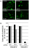
Figure 2. Role of B12 and B14 in formation of DJANGOs.
A. Images are individual confocal slices of HeLa cells stained with anti-HA to detect HA-tagged B12 or B14. The left panels show cells expressing an shRNA targeting an irrelevant gene (shLib1), the right panels show cells expressing an shRNA targeting B14 (top) or B12 (bottom). The top panels show cells over-expressing B12-HA while the bottom panels show cells over-expressing B14-HA. B. Random microscopic fields of cells treated as in panel A were scored for the presence of DJANGOS in two independent experiments (shown in black and grey bars) and normalized to the values for cells expressing Lib1 control shRNA. In these experiments, B12 induced DJANGOS in approximately 29% of control cells, and B14 induced DJANGOS in approximately 11% of control cells. The data were subjected to an F-test followed by an unequal variance t-test. B12 knock-down significantly reduced the ability of B14 to induce DJANGOS as compared to cells expressing the control shRNA (p<0.021), whereas B14 knockdown did not affect the ability of B12 to induce DJANGOS (p <0.8).
Figure 3
Figure 3. Co-localization of B12 with Hsc70 and other proteins in DJANGOS.
HeLa cells over-expressing B12-HA were stained with anti-B12 (A–F) or anti-HA (G) to detect B12 (in green) and with antibodies specific for the indicated cellular protein (in red). Overlapping signals in the merged images on the right are shown in yellow. The same confocal slice is shown in each row of panels. A. Hsc70. B. BiP. C. Sec61γ. D. Emerin. This section is from the same nucleus shown in Figure 1B. E. Lamin A. F. Lamin B. G. NPC. This slice is near the bottom of the nucleus.
Figure 4
Figure 4. Ultrastructure of DJANGOS in HeLa cells.
A. A low power electron micrograph of a cell over-expressing B12-HA shows multiple DJANGOS, labeled “D,” regularly spaced underneath the nuclear envelope. The dark granular objects labeled “nu” are nucleoli. B. Image of a cell over-expressing B12-HA and E2 shows intranuclear, tightly clustered, single-walled membrane tubes. C. Worm-like membrane tubes seem to transition from the cross-sectioned tubes in DJANGOS from cells over-expressing B12-HA. D. Cells expressing B14-HA and E2 also show intranuclear DJANGOS. The nuclear envelope and cytoplasm are visible in the lower right. E. Cells expressing B12-tGFP and E2 display concentric multi-lamellar nuclear whorls in close apposition to simple tubes. F. Complex membranous structure in cells expressing B12-tGFP and E2. G. Cytoplasmic and nuclear concentric multi-lamellar whorls in cells expressing B12-tGFP and E2. H. High-power EM images of cells over-expressing B12-HA stained with anti-BiP antibody and gold bead-conjugated secondary antibody. Membranes appear as a negative image, and the black spots are gold beads indicating sites of BiP reactivity. “N” and “C” indicate nucleus and cytoplasm, respectively.
Figure 5
Figure 5. DJANGOS emerge from atypical nuclear pores.
HeLa cells over-expressing B12-HA were visualized by electron microscopy. A. A double-walled membrane body within the nucleus connects to a tube (arrow), which then intermingles with other tubes in various orientations. The cytoplasm is at the lower left. B. Double-membrane body in proximity to the nuclear envelope connects to a complex structure extending into the nucleus. Cytoplasm is at left. C. Simple double-membrane structure extending into the nucleus from an atypical nuclear pore (indicated by the arrow). Cytoplasm is at top. D. Complex double-membrane structure containing two atypical nuclear pores (indicated by the arrows) connected to tubular structures inside the nucleus. Cytoplasm is at top.
Figure 6
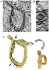
Figure 6. Electron tomography of DJANGOS.
A. Tomographic slice of an EM thick section showing a nuclear pore-associated structure formed in HeLa cells expressing B12-HA. Cytoplasm and nucleus are labeled with “C” and “N,” respectively. The thick arrow points to an atypical nuclear pore structure in cross-section at the junction with the nuclear envelope; the thin arrow points to a classic nuclear pore in the double membrane inside the nucleus. B. End-on view of the atypical nuclear pore at the neck of the double-membrane structure. C. 3D rendering of the tomogram shows the NPCs in blue. Also see Movie S1.
Figure 7
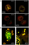
Figure 7. Live-cell imaging of DJANGOS.
Static images retrieved from live cell imaging of HeLaM or CV1 cells co-transfected with plasmids expressing B12-mCherry and LBR-eGFP. A. Top panels show two images of a HeLaM cell separated by 15 minutes acquired shortly before mitosis. Bottom panels shows images acquired one hour apart of the daughter cells of the cell shown in the top panels. See Movie S3. B. Left panel shows cytoplasmic billowing of B12 structures in CV1 cells; right panel shows cytoplasmic B12 “balloons on strings” in CV1 cells. See Movies S4 and S5.
Figure 8
Figure 8. DJANGOS formation requires DNAJ/Hsc70 activity.
A. RIPA buffer extracts were prepared from unmodified HeLa cells (H) or cells transduced with genes encoding wild-type B12-HA (B12) or the H138Q-HA mutant (12Q). Extracts were immunoprecipitated with anti-B12 and subjected to SDS-polyacrylamide gel electrophoresis, or electrophoresed without immunoprecipitation (lane 1). Top and bottom panels show anti-HA and Hsc70 immunoblots, respectively. B. HeLa cells were infected with retroviruses expressing the indicated wild-type or mutant B12-HA (top panels) or B14-HA (bottom panels) gene and stained with anti-HA to detect B12 or B14. Images are individual confocal slices. The left panels show cells expressing wild-type B12 or B14, the middle panels the J-domain mutants and the right panel the carboxy- truncated B12ΔC.

Similar articles

Cited by

References

    1. Imamoto N, Funakoshi T (2012) Nuclear pore dynamics during the cell cycle. Curr Opin Cell Biol 24: 453–459. - PubMed
    1. Schooley A, Vollmer B, Antonin W (2012) Building a nuclear envelope at the end of mitosis: coordinating membrane reorganization, nuclear pore complex assembly, and chromatin de-condensation. Chromosoma 121: 539–554. - PMC - PubMed
    1. Gruenbaum Y, Goldman RD, Meyuhas R, Mills E, Margalit A, et al. (2003) The nuclear lamina and its functions in the nucleus. Int Rev Cytol 226: 1–62. - PubMed
    1. Cronshaw JM, Krutchinsky AN, Zhang W, Chait BT, Matunis MJ (2002) Proteomic analysis of the mammalian nuclear pore complex. J Cell Biol 158: 915–927. - PMC - PubMed
    1. Adams RL, Wente SR (2013) Uncovering nuclear pore complexity with innovation. Cell 152: 1218–1221. - PMC - PubMed

Publication types

LinkOut - more resources