

Evaluation of inferior alveolar nerve regeneration by bifocal distraction osteogenesis with retrograde transportation of horseradish peroxidase in dogs
- PMID: 24732938
- PMCID: PMC3986082
- DOI: 10.1371/journal.pone.0094365
Evaluation of inferior alveolar nerve regeneration by bifocal distraction osteogenesis with retrograde transportation of horseradish peroxidase in dogs
Abstract
Background: Bifocal distraction osteogenesis has been shown to be a reliable method for reconstructing segmental mandibular defects. However, there are few reports regarding the occurrence of inferior alveolar nerve regeneration during the process of distraction. Previously, we reported inferior alveolar nerve regeneration after distraction, and evaluated the regenerated nerve using histological and electrophysiological methods. In the present study, we investigated axons regenerated by bifocal distraction osteogenesis using retrograde transportation of horseradish peroxidase in the mandibles of dogs to determine their type and function.
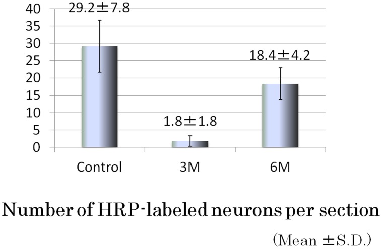
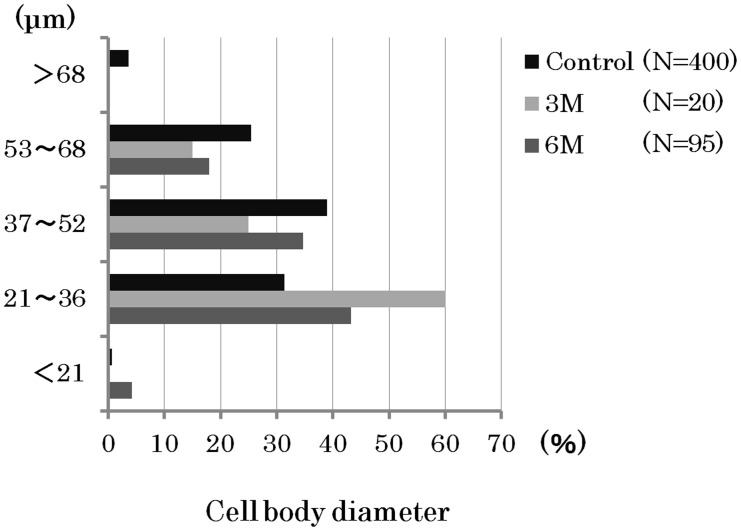
Methods and findings: Using a bifocal distraction osteogenesis method, we produced a 10-mm mandibular defect, including a nerve defect, in 11 dogs and distracted using a transport disk at a rate of 1 mm/day. The regenerated inferior alveolar nerve was evaluated by retrograde transportation of HRP in all dogs at 3 and 6 months after the first operation. At 3 and 6 months, HRP-labeled neurons were observed in the trigeminal ganglion. The number of HRP-labeled neurons in each section increased, while the cell body diameter of HRP-labeled neurons was reduced over time.
Conclusions: We found that the inferior alveolar nerve after bifocal distraction osteogenesis successfully recovered until peripheral tissue began to function. Although our research is still at the stage of animal experiments, it is considered that it will be possible to apply this method in the future to humans who have the mandibular defects.
Conflict of interest statement
Figures


Similar articles
-
Electrophysiologic evaluation of inferior alveolar nerve regenerated by bifocal distraction osteogenesis in dogs.Plast Reconstr Surg. 2013 Oct;132(4):877-882. doi: 10.1097/PRS.0b013e31829fe49a. Plast Reconstr Surg. 2013. PMID: 24076681
-
Inferior alveolar nerve regeneration after bifocal distraction osteogenesis in dogs.J Oral Maxillofac Surg. 2013 Oct;71(10):1810.e1-11. doi: 10.1016/j.joms.2013.04.037. Epub 2013 Jul 17. J Oral Maxillofac Surg. 2013. PMID: 23871317
-
Effect of nerve growth factor and Schwann cells on axon regeneration of distracted inferior alveolar nerve following mandibular lengthening.Chin J Traumatol. 2004 Apr;7(2):81-6. Chin J Traumatol. 2004. PMID: 15294125
-
Effects of mandibular distraction osteogenesis on the inferior alveolar nerve: an experimental study in monkeys.Plast Reconstr Surg. 2002 Jun;109(7):2373-83. doi: 10.1097/00006534-200206000-00032. Plast Reconstr Surg. 2002. PMID: 12045565
-
Alveolar distraction osteogenesis for dental implant treatments of the vertical bone atrophy: A systematic review.Med Oral Patol Oral Cir Bucal. 2019 Jan 1;24(1):e70-e75. doi: 10.4317/medoral.22750. Med Oral Patol Oral Cir Bucal. 2019. PMID: 30573711 Free PMC article.
References
-
- Costatino PD, Shybut G, Friedmann CD, Pelzer HJ, Masini M, et al. (1990) Segmental mandibular regeneration by distraction osteogenesis. Arch Otolaryngol Head Neck Surg 116: 535–545. - PubMed
-
- Costatino PD, Friedmann CD, Shindo ML, Houston G, Sisson GA Sr (1993) Experimental mandibular regrowth by distraction osteogenesis: Long-term results. Arch Otolaryngol Head Neck Surg 119: 511–516. - PubMed
-
- Ganey TM, Klotch DW, Slater-Haase AS, Sasse J (1994) Evaluation of distraction osteogenesis by scanning electron microscopy. Otolaryngol Head Neck Surg 111: 265–272. - PubMed
-
- Ganey TM, Klotch DW, Sasse J, Ogden JA, Garcia T (1994) Basement membrane of blood vessel during distraction osteogenesis. Clin Orthop Relat Res 301: 132–138. - PubMed
-
- Gantous A, Phillips JH, Catton P, Holmberg D (1994) Distraction osteogenesis in the irradiated canine mandible. Plast Reconstr Surg 93: 164–168. - PubMed
Publication types
MeSH terms
Substances
LinkOut - more resources
Full Text Sources
Other Literature Sources
