Review
. 2014 Jun;15(6):367-78.
doi: 10.1038/nrg3687. Epub 2014 Apr 15.

X chromosome regulation: diverse patterns in development, tissues and disease

Affiliations
Review

X chromosome regulation: diverse patterns in development, tissues and disease

Xinxian Deng et al. Nat Rev Genet. 2014 Jun.

Abstract

Genes on the mammalian X chromosome are present in one copy in males and two copies in females. The complex mechanisms that regulate the X chromosome lead to evolutionary and physiological variability in gene expression between species, the sexes, individuals, developmental stages, tissues and cell types. In early development, delayed and incomplete X chromosome inactivation (XCI) in some species causes variability in gene expression. Additional diversity stems from escape from XCI and from mosaicism or XCI skewing in females. This causes sex-specific differences that manifest as differential gene expression and associated phenotypes. Furthermore, the complexity and diversity of X dosage regulation affect the severity of diseases caused by X-linked mutations.


Conflict of interest statement

Competing interests statement

The authors declare no competing interests.

Figures

Figure 1
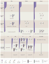
Figure 1. Variable gene content and dosage upregulation of the mammalian X chromosome
a | Schematics of the human and mouse X chromosomes, are shown. The X conserved region (XCR; blue), which occupies the long arm and a small region of the short arm of the human X chromosome, and the X added region (XAR; green) are indicated. In mice, the XAR and XCR are rearranged. Many of the newly acquired ampliconic genes differ between humans (pink) and mice (orange). Note that the position of these regions does not necessarily represent the position of the ampliconic genes. b | The proto-X and proto-Y chromosomes originally had similar sizes, and their expression level was balanced with that of autosomal genes (which are present in two copies). After the Y chromosome degenerated, hemizygous X-linked genes became upregulated. There is evidence of at least two feedforward mechanisms, which are schematically represented by the different colour faucets for upregulation of X-linked genes in mammals, including increased transcription (yellow and green; large grey arrows) and extended RNA half-life (pink and purple; large black arrows). Additional feedback and buffering mechanisms that respond to any type of aneuploidy may also be implicated in X upregulation (blue; small black arrows). Genes that represent dosage-insensitive genes (black) would not be upregulated. Note that some genes may be regulated by more than one mechanism (red and orange). Part b is adapted from REF. .
Figure 2
Figure 2. XCI initiation varies in mammals
A comparison of X chromosome inactivation (XCI) in early embryonic development (that is, from zygote to epiblast) in three eutherian species (mice, rabbits and humans) is shown. XCI is mainly mediated by regulation of X inactive specific transcript (XIST) in eutherians; high expression levels of XIST start at zygotic genome activation (ZGA), and mice have the earliest onset of Xist expression. In mice, XCI is initially imprinted and triggered by exclusive Xist expression and coating of the paternal X chromosome (Xp) at the 2–4-cell stage. At the morula stage, imprinted XCI is completed. The Xp remains inactive in the trophectoderm (TE) that develops into extra-embryonic tissues such as the placenta, whereas Xp is reactivated in the inner cell mass (ICM) of the mid-stage blastocyst. XCI is then reinitiated by upregulating Xist from one randomly chosen X chromosome (that is, either the maternal X chromosome (Xm) or Xp) at the late-stage blastocyst and epiblast stage. In rabbits, XIST is not imprinted; thus, XIST upregulation and coating can occur on one X chromosome or both X chromosomes in some cells at the morula and early blastocyst stages followed by silencing. In late-stage blastocysts, cells with random monoallelic XCI in both the ICM and TE are selected by an unknown mechanism. In humans, XIST is also not imprinted, and diffuse XIST coating is visible on both X chromosomes but without initiation of silencing. Random XCI is initiated much later, probably after the blastocyst stage. Pluripotency regulatory factors such as octamer-binding protein 4 (OCT4) can repress XIST. OCT4 expression is very high in zygotes, decreases sharply at the two-cell stage and then gradually declines until ZGA, followed by an increase in preimplantation embryos. OCT4 levels remain high in both the ICM and TE in rabbit and human blastocysts but only in the ICM in mice. Data from REF. .
Figure 3
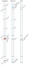
Figure 3. Escape from XCI varies between cell types and tissue in mice
Schematics show the location of genes that escape X chromosome inactivation (XCI) along the mouse X chromosome on the basis of complete surveys of allele-specific gene expression by RNA sequencing in three systems: trophoblasts, Patski cells (which are derived from the embryonic kidney) and the brain,,. The positions of five escape genes on the mouse X chromosome in all three cell types and tissue (black) are indicated, with their names shown at the left. Genes that escape XCI only in trophoblasts are shown in red, those only in Patski cells in blue and those only in the mouse brain in green; the gene names are indicated at the right of each chromosome. Only genes expressed from the inactive X chromosome at a level ≥10% of the active X chromosome in Patski cells (13 genes) and in the brain (8 genes), and genes with P < 0.05 in trophoblasts (16 genes; common in both reciprocal crosses) are included. Escape gene location is based on coordinates from University of California Santa Cruz (UCSC) genome browser build NCBI37/mm9.
Figure 4
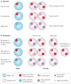
Figure 4. Variability of X-linked gene expression and sex bias
a | Sex bias in X-linked gene expression in normal individuals is shown. There is no sex bias for genes that are subject to X chromosome inactivation (XCI) or for genes with equivalent X–Y paralogues (not shown). A female bias occurs for genes that escape XCI but that have no equivalent Y paralogue; a male bias occurs for genes that are solely expressed in male organs such as the testes. b | The effects of X-linked gene mutation depend on XCI patterns. For genes that are subject to XCI, a mutation that affects males does not necessarily affect females, who can be rescued either by random XCI (which leads to mosaicism for cells with the mutant allele silenced) or by selective skewing in favour of cells that express the normal allele. For escape genes, males can be partly rescued by expression of a Y paralogue with similar function (if it exists), whereas females will be haploinsufficient even if there is skewing in favour of the normal allele. Severity of the haploinsufficiency in females depends on the expression levels from the inactive X chromosome. Mutations in genes that are essential for XCI (such as the gene that encodes X inactive specific transcript (XIST)) specifically affect females by causing XCI failure. If a mutation is in an X-linked gene that is solely expressed in male organs (such as the testes), only males will be affected. However, as many of the testis-specific genes are ampliconic, the effect of a mutation in one copy will be partly rescued by the normal copies.

References

    1. Livernois AM, Graves JA, Waters PD. The origin and evolution of vertebrate sex chromosomes and dosage compensation. Hered (Edinb) 2012;108:50–58. - PMC - PubMed
    1. Disteche CM. Dosage compensation of the sex chromosomes. Annu Rev Genet. 2012;46:537–560. - PMC - PubMed
    1. Bachtrog D. Y-chromosome evolution: emerging insights into processes of Y-chromosome degeneration. Nature Rev Genet. 2013;14:113–124. - PMC - PubMed
    1. Gurbich TA, Bachtrog D. Gene content evolution on the X chromosome. Curr Opin Genet Dev. 2008;18:493–498. - PMC - PubMed
    1. Wang PJ, McCarrey JR, Yang F, Page DC. An abundance of X-linked genes expressed in spermatogonia. Nature Genet. 2001;27:422–426. - PubMed

Publication types

Supplementary concepts