. 2014 Jul;82(7):2713-27.
doi: 10.1128/IAI.01600-13. Epub 2014 Apr 14.

Site-dependent recruitment of inflammatory cells determines the effective dose of Leishmania major

Affiliations

Site-dependent recruitment of inflammatory cells determines the effective dose of Leishmania major

Flavia L Ribeiro-Gomes et al. Infect Immun. 2014 Jul.

Abstract

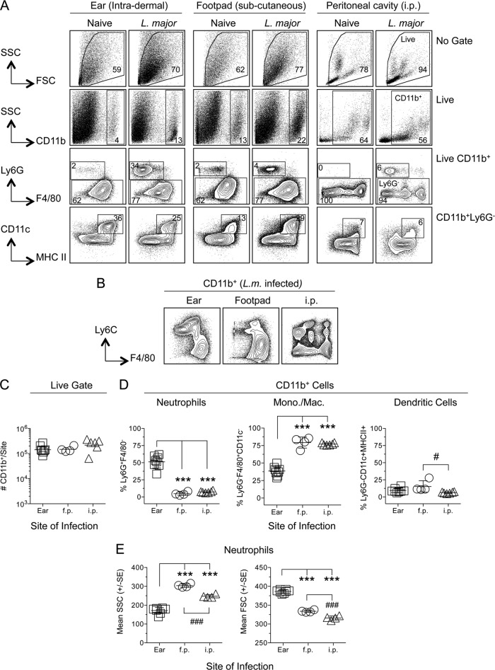
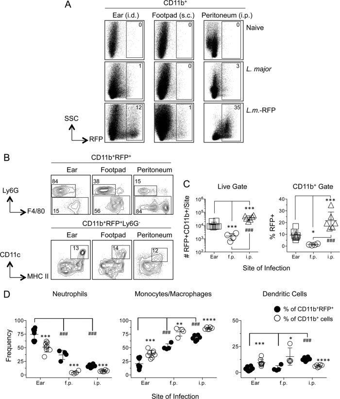
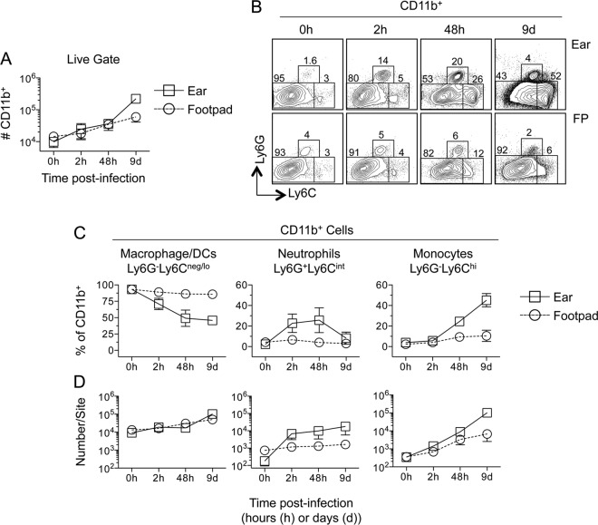
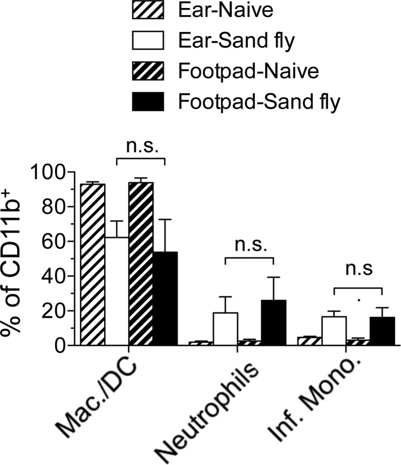
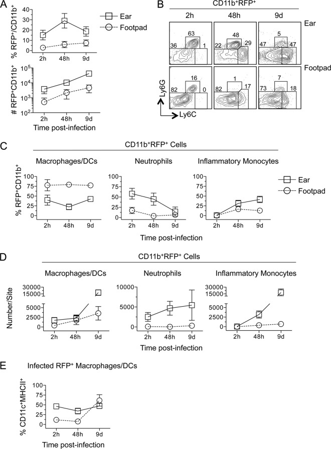
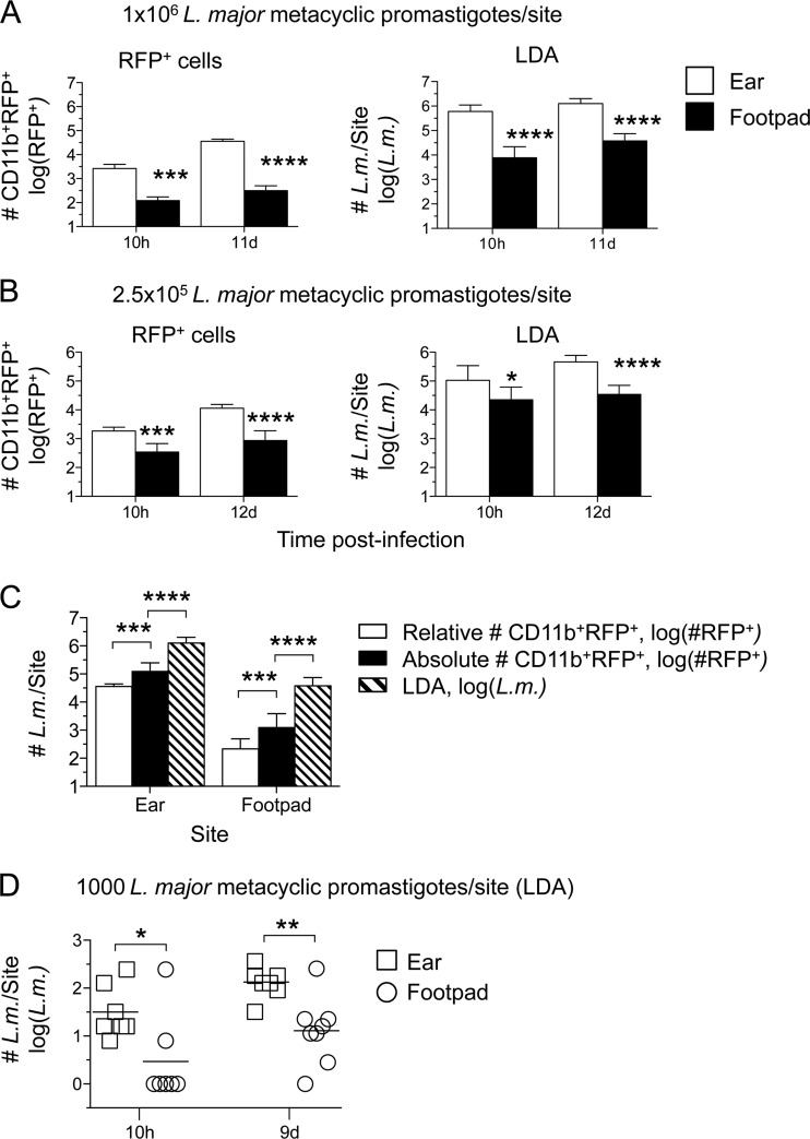
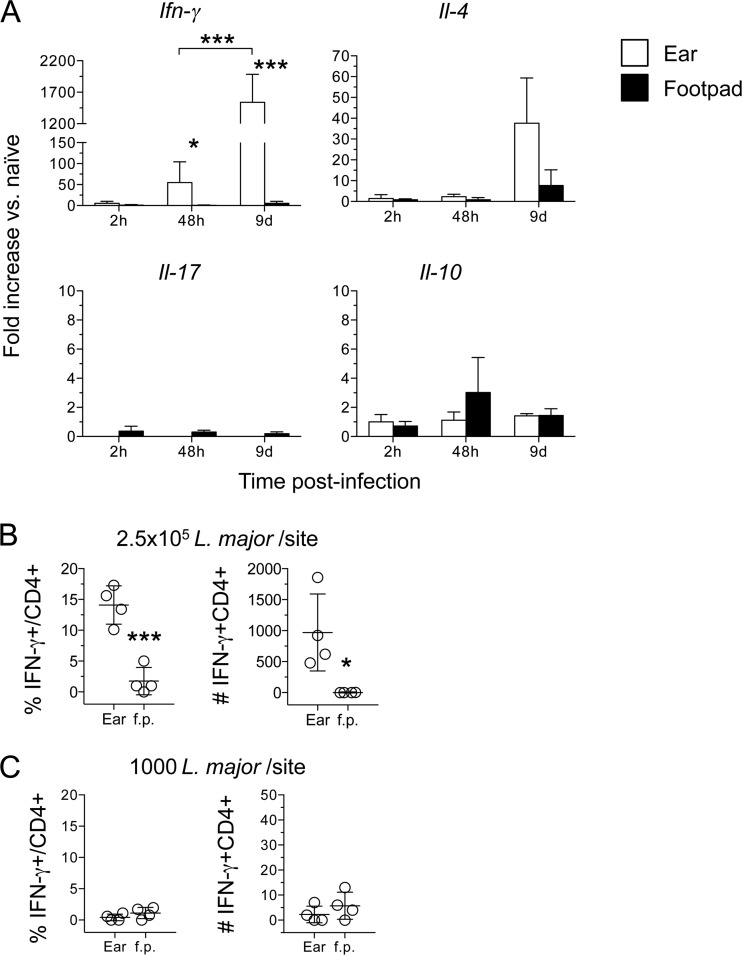
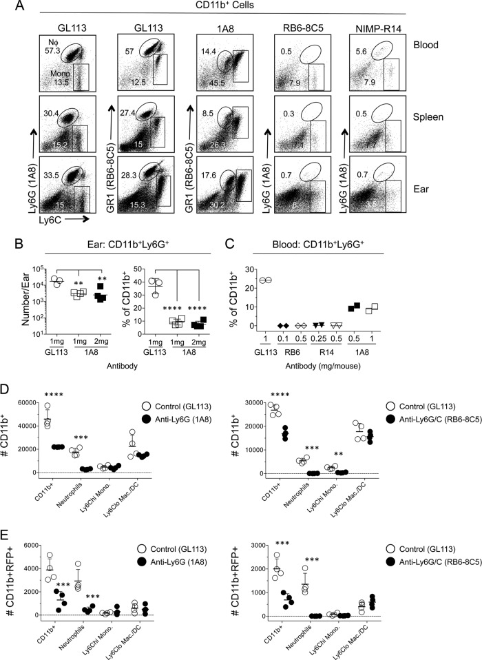
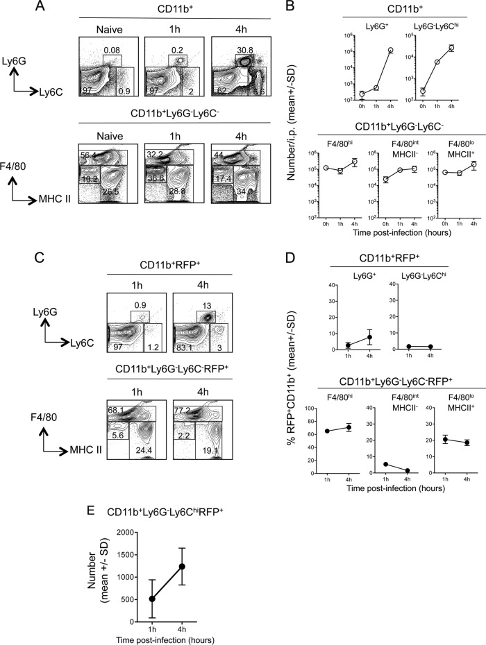
The route of pathogen inoculation by needle has been shown to influence the outcome of infection. Employing needle inoculation of the obligately intracellular parasite Leishmania major, which is transmitted in nature following intradermal (i.d.) deposition of parasites by the bite of an infected sand fly, we identified differences in the preexisting and acute cellular responses in mice following i.d. inoculation of the ear, subcutaneous (s.c.) inoculation of the footpad, or inoculation of the peritoneal cavity (intraperitoneal [i.p.] inoculation). Initiation of infection at different sites was associated with different phagocytic populations. Neutrophils were the dominant infected cells following i.d., but not s.c. or i.p., inoculation. Inoculation of the ear dermis resulted in higher frequencies of total and infected neutrophils than inoculation of the footpad, and these higher frequencies were associated with a 10-fold increase in early parasite loads. Following inoculation of the ear in the absence of neutrophils, parasite phagocytosis by other cell types did not increase, and fewer parasites were able to establish infection. The frequency of infected neutrophils within the total infected CD11b(+) population was higher than the frequency of total neutrophils within the total CD11b(+) population, demonstrating that neutrophils are overrepresented as a proportion of infected cells. Employing i.d. inoculation to model sand fly transmission of parasites has significant consequences for infection outcome relative to that of s.c. or i.p. inoculation, including the phenotype of infected cells and the number of parasites that establish infection. Vector-borne infections initiated in the dermis likely involve adaptations to this unique microenvironment. Bypassing or altering this initial step has significant consequences for infection.


Figures

FIG 1
FIG 1
Phenotypic analysis of phagocytic cells following needle inoculation of the ear (intradermal), footpad (f.p.) (subcutaneous), or peritoneal cavity (i.p.) with L. major-RFP reveals site-specific populations of CD11b+ cells. Individual C57BL/6 mice were injected with 1 × 10⁶ L. major-RFP metacyclic promastigotes via the indicated route. Ten hours postinfection, cells from naïve or infected mice were prepared, stained for the indicated surface markers, and analyzed by flow cytometry. (A) Representative dot plots and gating strategy of phagocytic populations. (B) Equal portions of the individual samples for which results are reported in panels C and D were pooled, and the pooled samples were stained for the indicated surface markers and analyzed by flow cytometry. (C) Total relative number of CD11b+ cells recovered from each site of analysis. Results for 8 (ear), 4 (f.p.), or 6 (i.p.) mice are shown. (D) Percentages of neutrophils (CD11b+ Ly6G+ F4/80), monocytes/macrophages (CD11b+ Ly6G [MHC-II CD11c]), or dendritic cells (CD11b+ Ly6G [MHC-II CD11c]+) among total CD11b+ cells. A log scale was employed here and elsewhere in order to display large differences in cell numbers accurately. (E) SSC and FSC characteristics of CD11b+ F4/80 Ly6G+ neutrophils derived from the ear, f.p., or peritoneal cavity 2 h following inoculation with 1 × 10⁶ L. major parasites. Horizontal lines represent the means ± SD (C and D) or standard errors (E). Asterisks indicate a significant difference from the ear group; number signs indicate a significant difference from the f.p. group. The levels of significance represented by various numbers of asterisks are explained in Materials and Methods. Data are representative of 2 independent experiments.
FIG 2
FIG 2
Phenotypic analysis of L. major-infected CD11b+ RFP+ cells in the ear (i.d.), footpad (s.c.), and peritoneal cavity reveals increased frequencies of infected neutrophils in the ear and preferential parasite capture by neutrophils. Samples for which results are shown in Fig. 1 were analyzed as a function of RFP expression. (A) Representative dot plots of RFP expression by CD11b+ cells in naïve mice or in mice injected with wild-type or RFP-expressing L. major parasites. (B) Representative dot plots and gating strategy of CD11b+ RFP+ cells. (C) Total relative number of CD11b+ RFP+ cells (left) or frequency of RFP+ cells within the CD11b+ gate (right) as a function of site. (D) Frequencies of the indicated myeloid populations among total CD11b+ RFP+ cells (filled circles) or total CD11b+ cells (open circles). Asterisks indicate a significant difference between total CD11b+ cells and CD11b+ RFP+ cells. Number signs indicate a significant difference between groups as indicated by the brackets. Horizontal lines represent means ± SD. Data are representative of 2 independent experiments.
FIG 3
FIG 3
Kinetic analysis of myeloid cell recruitment reveals enhanced recruitment to the ear as opposed to the footpad following the inoculation of L. major-RFP. Mice were injected with 2.5 × 10⁵ L. major-RFP parasites i.d. in the ear or s.c. in the footpad. At the indicated time points p.i., each site was analyzed for the indicated myeloid populations. (A) Total relative numbers of CD11b+ cells at each site. d, day. (B) Representative dot plots of Ly6G and Ly6C expression on CD11b+ cells. (C and D) Frequencies of the indicated myeloid populations among CD11b+ cells (C) or total relative number of each population (D) at each site over the indicated time p.i. Data are means ± SD for 5 to 6 (ear) or 5 to 7 (f.p.) samples. Data are representative of 2 independent experiments.
FIG 4
FIG 4
Sand fly bites elicit equivalent recruitment of inflammatory cells to the footpad or ear skin. Shown are the frequencies of Ly6G Ly6Cneg/lo macrophages/DCs, CD11b+ Ly6G+ Ly6Cintermediate neutrophils, or Ly6G Ly6Chi inflammatory (Inf.) monocytes among CD11b+ cells 10 h following exposure to the bites of uninfected P. duboscqi sand flies. Data are means ± SD for 11 to 12 (sand fly-exposed ears or footpads) or 4 (naïve ears or footpads) samples. Data are pooled from 2 independent experiments with similar results. n.s., not significant.
FIG 5
FIG 5
Increased neutrophil recruitment correlates with increased numbers of RFP+ cells following i.d. inoculation of the ear versus s.c. inoculation of the footpad. Samples for which results are shown in Fig. 3 were analyzed as a function of RFP expression. (A) Frequency of infected (RFP+) CD11b+ cells among total CD11b+ cells (top) or total relative number of CD11b+ RFP+ cells at each site over the indicated time p.i. (B) Representative dot plots of Ly6G and Ly6C expression on CD11b+ RFP+ cells. (C and D) Frequencies of the indicated myeloid populations among CD11b+ RFP+ cells (C) or total relative number of each population (D) at each site over the indicated time p.i. (E) Frequency of CD11c+ MHC II+ cells within the infected CD11b+ Ly6G Ly6Clo RFP+ or CD11b+ Ly6G Ly6Chi RFP+ population at each site over time. Each symbol represents the mean, and error bars represent SD, for 5 to 6 (ear) or 5 to 7 (f.p.) samples. Data are representative of 2 independent experiments.
FIG 6
FIG 6
Intradermal inoculation of the ear results in a higher effective dose than s.c. inoculation of the footpad. Mice were infected i.d. in the ear or s.c. in the footpad with L. major-RFP. At the indicated time points, the number of CD11b+ RFP+ cells (RFP+ cells), or the number of parasites as determined by limiting dilution analysis (LDA), per ear or footpad was determined. Analysis was performed following needle inoculation of 1 × 10⁶ (A), 2.5 × 10⁵ (B), or 1,000 (D) metacyclic promastigotes. (C) Day 12 p.i. data from the experiment for which results are shown in panel A were plotted with the absolute number of CD11b+ RFP+ cells per site as determined by employing counting beads. Six to 12 ears or footpads were analyzed per time point. Asterisks indicate a significant difference between the ear and footpad groups (A, B, and D) or between the groups indicated by brackets (C). (D) *, P = 0.012; **, P = 0.008.
FIG 7
FIG 7
Decreased parasite numbers following s.c. inoculation of the footpad are not due to earlier onset of adaptive immunity. (A) Total RNA was isolated directly from the ears or footpads for which results are shown in Fig. 3, reverse transcribed, and analyzed by real-time PCR. The expression of the target genes was normalized to that of an endogenous control, and the values shown are fold increases over expression in naïve ears or footpads. Data are means ± SD for 3 to 5 samples. (B and C) Intracellular staining for IFN-γ. The CD3+ CD4+ T cells used were from ear or footpad samples for which results are shown in Fig. 6B, day 12 (B), or in Fig. 6D, day 9 (C). Asterisks indicate a significant difference between the ear and footpad groups or between the groups indicated by brackets. Data are means ± SD for 4 samples.
FIG 8
FIG 8
A significant portion of needle-inoculated parasites fail to establish infection in the absence of neutrophils. (A to E) Mice were injected i.p. with antibody 1A8, RB6-8C5, or NIMP-R14 prior to needle inoculation of the ear with 7.5 × 10⁵ to 1 × 10⁶ L. major metacyclic-promastigotes. Sixteen hours p.i., CD11b+ cells from the ear, spleen, and blood were assessed for CD11b, Ly6G, and Ly6C expression. (A) Representative dot plots of Ly6C and Ly6G expression on CD11b+ cells from mice treated with 0.5 mg of antibody 24 h prior to L. major inoculation. (B) Total relative number (left) or frequency (right) of CD11b+ Ly6G+ cells in the ears of mice treated with Ab 1A8. Mice received either a single 1-mg dose of 1A8 24 h prior to challenge or two 1-mg doses of 1A8, one at 24 h and one at 48 h prior to challenge. Horizontal lines indicate means ± SD for 3 to 4 mice per experiment. (C) Percentages of CD11b+ Ly6G+ neutrophils in the blood of mice treated with the indicated doses of antibodies 24 h prior to challenge. (D and E) Mice were treated with the antibody 1A8 (left) or RB6-8C5 (right) 24 h prior to injection with L. major. Ears were analyzed for the total relative numbers of the indicated CD11b+ (D) or CD11b+ RFP+ (E) myeloid populations. Horizontal lines represent means ± SD for 4 mice. Data are representative of 2 independent experiments. Asterisks indicate a significant difference between groups as indicated by the brackets (B) or between GL113 and IA8- or RB6-8C5-treated mice for each cellular phenotype (D and E). Similar data were obtained by employing 0.5 or 1 mg of antibody.
FIG 9
FIG 9
Analysis of total and infected CD11b+ myeloid populations in the peritoneal cavity reveals that inflammatory monocytes are rapidly recruited but phagocytose relatively few parasites. Mice were injected i.p. with 2 × 10⁶ L. major-RFP parasites, and at the indicated time points p.i., myeloid cells were analyzed for RFP expression. (A) Representative dot plots and gating strategy of CD11b+ cells. (B) Total relative numbers of the indicated myeloid populations per cavity. Results for 3 mice are shown. (C) Representative dot plots and gating strategy of CD11b+ RFP+ cells. (D) Frequencies of the indicated myeloid populations among total CD11b+ RFP+ cells. (E) Total relative numbers of infected (RFP+) inflammatory monocytes in the peritoneal cavity at 1 and 4 h p.i. Symbols represent means; error bars indicate SD. Data are representative of 2 independent experiments.

References

    1. Mahmoudzadeh-Niknam H, Khalili G, Abrishami F, Najafy A, Khaze V. 2013. The route of Leishmania tropica infection determines disease outcome and protection against Leishmania major in BALB/c mice. Korean J. Parasitol. 51:69–74. 10.3347/kjp.2013.51.1.69 - DOI - PMC - PubMed
    1. Oliveira DM, Costa MA, Chavez-Fumagalli MA, Valadares DG, Duarte MC, Costa LE, Martins VT, Gomes RF, Melo MN, Soto M, Tavares CA, Coelho EA. 2012. Evaluation of parasitological and immunological parameters of Leishmania chagasi infection in BALB/c mice using different doses and routes of inoculation of parasites. Parasitol. Res. 110:1277–1285. 10.1007/s00436-011-2628-5 - DOI - PubMed
    1. Tabbara KS, Peters NC, Afrin F, Mendez S, Bertholet S, Belkaid Y, Sacks DL. 2005. Conditions influencing the efficacy of vaccination with live organisms against Leishmania major infection. Infect. Immun. 73:4714–4722. 10.1128/IAI.73.8.4714-4722.2005 - DOI - PMC - PubMed
    1. Osorio Y, Melby PC, Pirmez C, Chandrasekar B, Guarin N, Travi BL. 2003. The site of cutaneous infection influences the immunological response and clinical outcome of hamsters infected with Leishmania panamensis. Parasite Immunol. 25:139–148. 10.1046/j.1365-3024.2003.00615.x - DOI - PubMed
    1. Baldwin TM, Elso C, Curtis J, Buckingham L, Handman E. 2003. The site of Leishmania major infection determines disease severity and immune responses. Infect. Immun. 71:6830–6834. 10.1128/IAI.71.12.6830-6834.2003 - DOI - PMC - PubMed

Publication types