. 2014 Apr 14;9(4):e94732.
doi: 10.1371/journal.pone.0094732. eCollection 2014.

Chymase mediates injury and mitochondrial damage in cardiomyocytes during acute ischemia/reperfusion in the dog

Affiliations

Chymase mediates injury and mitochondrial damage in cardiomyocytes during acute ischemia/reperfusion in the dog

Junying Zheng et al. PLoS One. .

Abstract

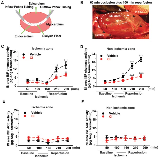
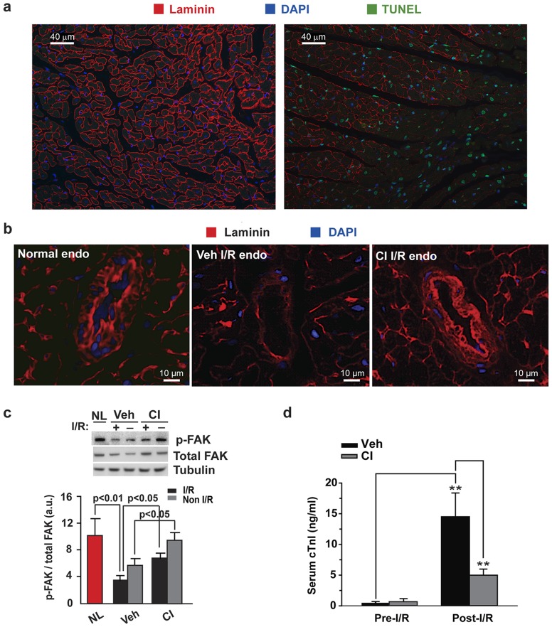
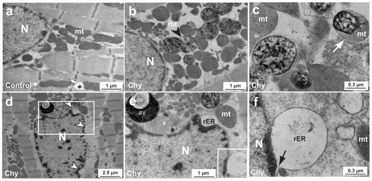
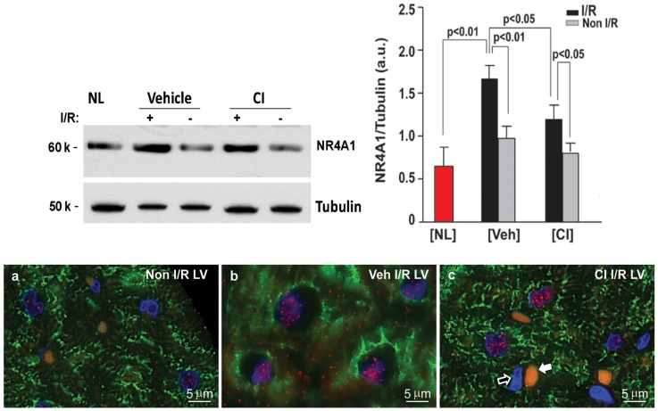
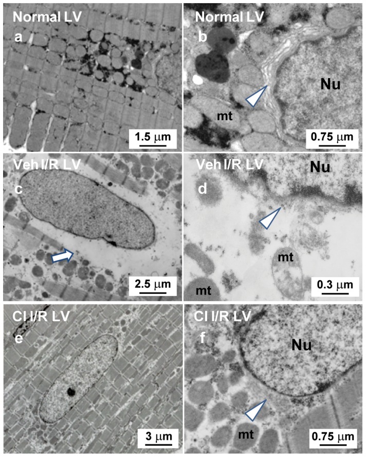
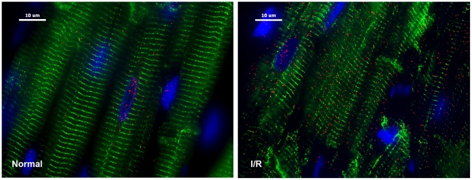
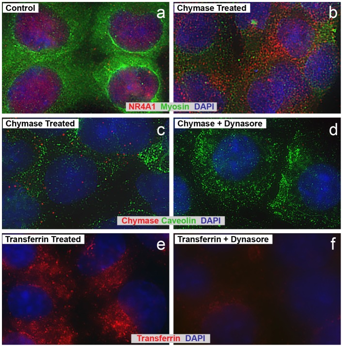
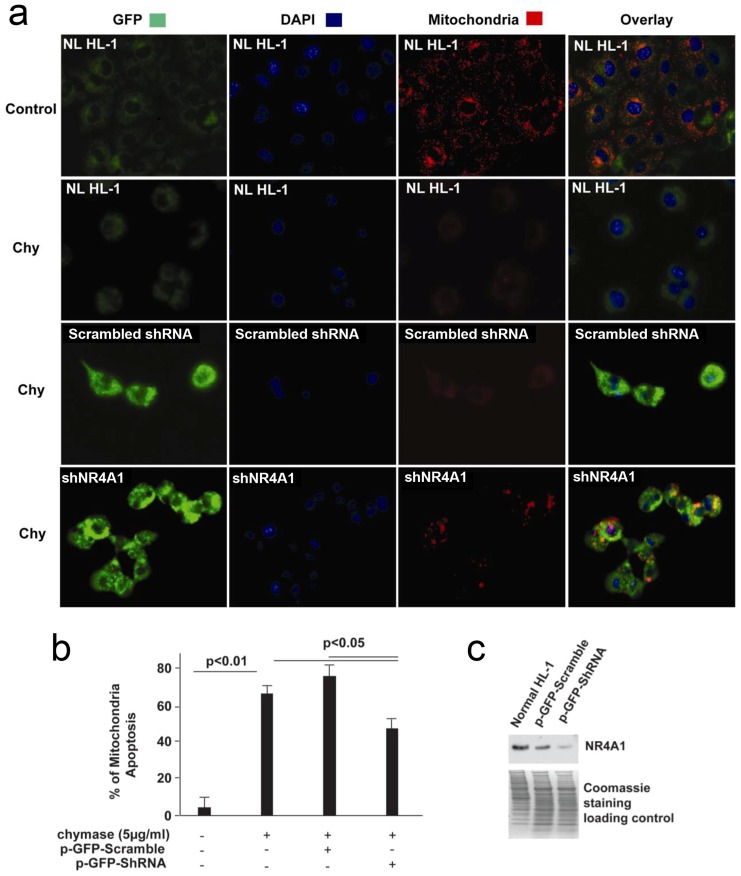
Cardiac ischemia and reperfusion (I/R) injury occurs because the acute increase in oxidative/inflammatory stress during reperfusion culminates in the death of cardiomyocytes. Currently, there is no drug utilized clinically that attenuates I/R injury in patients. Previous studies have demonstrated degranulation of mast cell contents into the interstitium after I/R. Using a dog model of I/R, we tested the role of chymase, a mast cell protease, in cardiomyocyte injury using a specific oral chymase inhibitor (CI). 15 adult mongrel dogs had left anterior descending artery occlusion for 60 min and reperfusion for 100 minutes. 9 dogs received vehicle and 6 were pretreated with a specific CI. In vivo cardiac microdialysis demonstrated a 3-fold increase in interstitial fluid chymase activity in I/R region that was significantly decreased by CI. CI pretreatment significantly attenuated loss of laminin, focal adhesion complex disruption, and release of troponin I into the circulation. Microarray analysis identified an I/R induced 17-fold increase in nuclear receptor subfamily 4A1 (NR4A1) and significantly decreased by CI. NR4A1 normally resides in the nucleus but can induce cell death on migration to the cytoplasm. I/R caused significant increase in NR4A1 protein expression and cytoplasmic translocation, and mitochondrial degradation, which were decreased by CI. Immunohistochemistry also revealed a high concentration of chymase within cardiomyocytes after I/R. In vitro, chymase added to culture HL-1 cardiomyocytes entered the cytoplasm and nucleus in a dynamin-dependent fashion, and promoted cytoplasmic translocation of NR4A1 protein. shRNA knockdown of NR4A1 on pre-treatment of HL-1 cells with CI significantly decreased chymase-induced cell death and mitochondrial damage. These results suggest that the beneficial effects of an orally active CI during I/R are mediated in the cardiac interstitium as well as within the cardiomyocyte due to a heretofore-unrecognized chymase entry into cardiomyocytes.


Conflict of interest statement

Competing Interests: The authors have declared that no competing interests exist.

Figures

Figure 1
Figure 1. In vivo cardiac microdialysis demonstrates increased ISF chymase activity during I/R that is decreased by pretreatment with chymase inhibitor (CI).
Panel a demonstrates the concept of cardiac microdialysis and panel b demonstrates the three microdialysis probes located in the area of I/R region (white arrows) and the two probes in the non-I/R region (black arrows) in the open chest dog heart. Panels c and d demonstrate ISF chymase activity in I/R zone (c) and non-I/R zone (d) in the Veh (black) and CI (red) pretreated dogs. Panels e and f demonstrate ISF ACE activity in the I/R (e) and non-I/R zones (f) in Veh and CI dogs. The vehicle chymase activity increased during reperfusion and therefore the analysis compared the vehicle baseline with increasing chymase activity during reperfusion. †† p<0.01 vehicle baseline vs. vehicle during ischemia;††† p<0.0001 vehicle baseline vs. vehicle during reperfusion; * p<0.05, *** p<0.001 vehicle vs. CI treated dogs. Veh dog, n = 9. CI dog, n = 6.
Figure 2
Figure 2. CI decreases laminin degradation and attenuates I/R injury in the dog heart.
(a) Compared to the normal LV (left panel), I/R results in a decrease in the laminin staining in this border region (right panel). There is a greater increase in apoptotic cells (green) with loss of laminin. (b) CI treatment decreased I/R induced laminin degradation. (c) FAK dephosphorylation is markedly decreased in normal (NL) dog LV compared to I/R LV. CI treatment attenuated I/R induced p-FAK dephosphorylation. (d) CI pretreatment significantly attenuates the I/R-induced release of cTnI into the circulation. ** p<0.01. NL dog, n = 3; Veh dog, n = 6; CI dog, n = 5.
Figure 3
Figure 3. Chymase promotes cell detachment and death in vitro in laminin-plated dog cardiomyocytes.
Control dog cardiomyocyte TEM is shown in the upper left panel (a) and images of chymase-treated cardiomyocytes (Chy) are shown in the other panels (b–f). Black arrowhead points to degraded mitochondria (mt, b). White arrow points to a partially degraded mitochondria (c). In bottom d–f panels, chymase is shown to induce the vacuolation of ER in the dog cardiomyocytes (white arrowheads, d). A blowup of the boxed area of the left panel (d) is shown in middle panel (e) and a larger blowup is shown in the right panel (f) further demonstrating the vacuolation of ER and autophagic vacuoles of degraded perinuclear mitochondria. Black arrow points to the membrane of ER and nucleus (f). mt, mitochondria; N, nucleus; rER, rough endoplasmic reticulum; av, autophagic vacuole.
Figure 4
Figure 4. Regulation of NR4A1 mRNA in normal, vehicle and CI treated dog LV I/R and non-I/R regions by microarray and RT-PCR.
Table (a) demonstrates Genesping GX.11 fold change from microarray and p value for mRNA intensity value of NR4A1 and ATF3 in normal, I/R and non-I/R vehicle- and CI-treated dog LV. Panel b demonstrates real-time RT-PCR validation of NR4A1 (left) and ATF3 (right) mRNA in normal, I/R and non-I/R vehicle- (Veh-nIR) and CI-treated dog LV. Normal dog, n = 5; Veh dog, n = 6; CI dog, n = 6.
Figure 5
Figure 5. NR4A1 protein expression in normal, vehicle- and CI-treated LV.
Upper panels show western blot and quantitation of NR4A1 in the I/R LV. The increase of NR4A1 protein in the I/R region is significantly attenuated by CI pretreatment. Lower panel a demonstrates very little NR4A1 (red) in the non-I/R LV region, while panel b demonstrates increased nuclear NR4A1 and its cytoplasmic migration in vehicle-treated I/R heart, but not in the CI-treated heart (panel c). Panel c also demonstrates the negative staining for NR4A1 in the endothelial cell nucleus (open arrow) that is located beside a red blood cell (white arrow). The yellowish color of red blood cell is caused by auto-fluorescence. Normal dog, n = 3; Veh dog, n = 6; CI dog, n = 5.
Figure 6
Figure 6. TEM demonstrate perinuclear degradation of endoplasmic reticulum (ER) and mitochondria degradation with I/R.
TEM images demonstrate tightly packed distribution of perinuclear mitochondria (a) and at higher magnification the location of ER (b, white arrow head). Middle panels (c and d) demonstrate the perinuclear vacuole that is also present by immunohistochemistry in Figure 3d–f, demonstrating breakdown of perinuclear mitochondria and ER (white arrow head). Lower panels (e and f) demonstrate that degradation of ER and mitochondria are prevented by CI pretreatment. Nu: nucleus; mt: mitochondria
Figure 7
Figure 7. Chymase inside dog cardiomyocytes during ischemia/reperfusion (I/R).
Adult dogs with 60 min of LAD occlusion and 100 min of reperfusion (right panel) and normal control (left panel). I/R LV demonstrates marked increase in chymase (red) with areas of breakdown of desmin (green, right) vs. normal control tissue (left). Blue: DAPI.
Figure 8
Figure 8. Chymase promotes cytoplasmic translocation of NR4A1 in HL-1 cells.
Immunocytochemistry demonstrates NR4A1 nuclear location at control (non-chymase treated) in HL-1 cells (panel a). Chymase (2.5 µg/ml) treatment for 2 h induces NR4A1 (red) cytoplasmic translocation in HL-1 cells as well as myosin (green) disruption (panel b). Active chymase enters HL-1 cells and is prevented by Dynasore (panel c and d). There is a small amount of chymase at control in HL-1 cells (not shown) and marked entry into HL-1 cell nuclei and cytoplasm after treatment with chymase (5 µg/ml) for 2 h that is prevented by pre-treatment with Dynasore. Lack of co-staining with caveolin 3 (green) demonstrates that chymase is not transported via caveolae. Dynasore prevents transferrin uptake in HL-1 cells. There is marked entry of transferrin into HL-1 cell nuclei and cytoplasm after treatment with transferrin (5 µg/ml, panel e) for 2 h that is prevented by pre-treatment with Dynasore (panel f).
Figure 9
Figure 9. Knockdown of NR4A1 significantly attenuates chymase-induced mitochondria (Mt) apoptosis.
(a) Immunofluorescence pattern of HL-1 cells transfected with p-GFP-shNR4A1 and scrambled p-GFP-shRNA for 5 days, loaded with dye (MitoCapture) and then treated with 5 µg/ml chymase (chy) for 2 h. Chymase treatment induces Mt apoptosis that is detected by loss of an electrochemical gradient and the inability to take up the MitoCapture dye. Normal Mt; bright red; Apoptotic Mt: little or no red staining. Bright green is the green fluorescent protein (GFP) and indicates that the cells were transfected with either p-GFP-shNR4A1 or scrambled p-GFP-shRNA. Cells without transfection and chymase treatment are used as the control. (b) Percentage of Mt apoptosis in chymase treated HL-1 cells, HL-1 cells transfected with p-GFP-shNR4A1 and scrambled p-GFP-shRNA. (c) NR4A1 protein expression in normal HL-1 cells, HL-1 cells transfected with p-GFP-shNR4A1 and scrambled p-GFP-shRNA. NL: HL-1 cells without transfection.

Similar articles

Cited by

References

    1. Yellon DM, Hausenloy DJ. Myocardial reperfusion injury (2007) N Engl J Med. 357: 1121–1135. - PubMed
    1. Frangogiannis NG, Perrard JL, Mendoza LH, Burns AR, Lindsey ML, et al. (1998) Stem cell factor induction is associated with mast cell accumulation after canine myocardial ischemia and reperfusion. Circulation 98: 687–698. - PubMed
    1. Wei CC, Hase N, Inoue Y, Bradley EW, Yahiro E, et al. (2010) Mast cell chymase limits the cardiac efficacy of Ang I-converting enzyme inhibitor therapy in rodents. J Clin Invest 120: 1229–1239. - PMC - PubMed
    1. Caughey GH, Raymond WW, Wolters PJ (2000) Angiotensin II generation by mast cell alpha- and beta-chymases. Biochim Biophys Acta 1480: 245–257. - PubMed
    1. Vartio T, Seppa H, Vaheri A (1981) Susceptibility of soluble and matrix fibronectins to degradation by tissue proteinases, mast cell chymase and cathepsin G. J Biol Chem. 256: 471–477. - PubMed

Publication types

MeSH terms