

Renal involvement in non-Hodgkin lymphoma: proven by renal biopsy
- PMID: 24733356
- PMCID: PMC3986362
- DOI: 10.1371/journal.pone.0095190
Renal involvement in non-Hodgkin lymphoma: proven by renal biopsy
Abstract
Aims: To determine the spectrum of renal lesions in patients with kidney involvement in non-Hodgkin's lymphoma (NHL) by renal biopsy.
Methods: The clinical features and histological findings at the time of the renal biopsy were assessed for each patient.
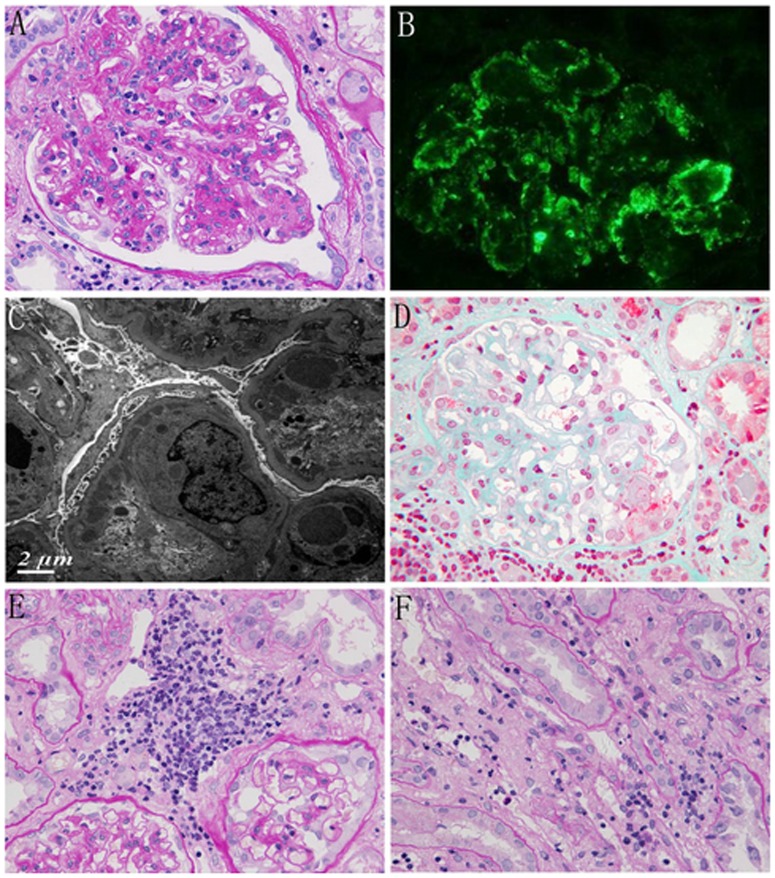
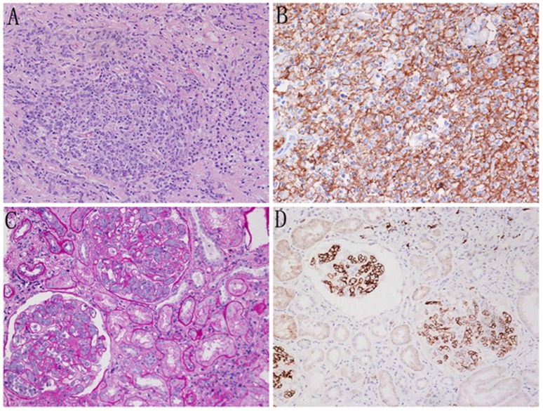
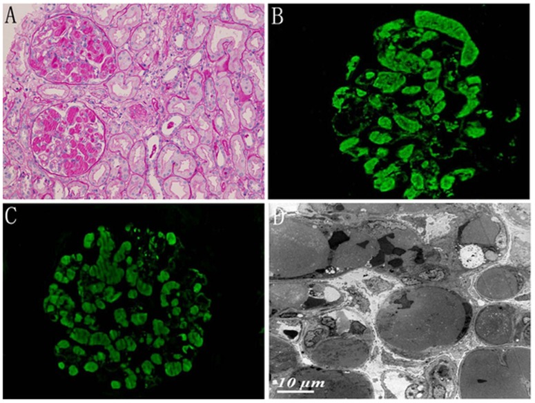
Results: We identified 20 patients with NHL and renal involvement, and the diagnosis of NHL was established following the kidney biopsy in 18 (90%) patients. The types of NHL include the following: chronic lymphocytic leukemia/small lymphocytic lymphoma (n = 8), diffuse large B-cell lymphoma (n = 4), T/NK cell lymphoma (n = 3), lymphoplasmacytic lymphoma (n = 2), cutaneous T-cell lymphoma (n = 1), mucosa-associated lymphoid tissue lymphoma (n = 1) and mantle cell lymphoma (n = 1). All presented with proteinuria, and 15 patients had impaired renal function. The pathological findings included (1) membranoproliferative glomerulonephritis-like pattern in seven patients; (2) crescent glomerulonephritis in four; (3) minimal-change disease in three, and glomeruli without specific pathological abnormalities in three; (4) intraglomerular large B-cell lymphoma in one; (5) intracapillary monoclonal IgM deposits in one; (6) primary diffuse large B-cell lymphoma of the kidneys in one; and (7) lymphoma infiltration of the kidney in eight patients.
Conclusion: A wide spectrum of renal lesions can be observed in patients with NHL, and NHL may be first proven by renal biopsies for evaluation of kidney injury or proteinuria. Renal biopsy is necessary to establish the underlying cause of renal involvement in NHL.
Conflict of interest statement
Figures
Similar articles
-
Patterns of glomerular injury in kidneys infiltrated by lymphoplasmacytic neoplasms.Hum Pathol. 2011 Jun;42(6):896-903. doi: 10.1016/j.humpath.2010.09.009. Epub 2011 Feb 1. Hum Pathol. 2011. PMID: 21288559
-
Clinicopathological features and individualized treatment of kidney involvement in B-cell lymphoproliferative disorder.Front Immunol. 2022 Sep 12;13:903315. doi: 10.3389/fimmu.2022.903315. eCollection 2022. Front Immunol. 2022. PMID: 36172352 Free PMC article.
-
Kidney involvement and renal manifestations in non-Hodgkin's lymphoma and lymphocytic leukemia: a retrospective study in 700 patients.Eur J Haematol. 2001 Sep;67(3):158-64. doi: 10.1034/j.1600-0609.2001.5790493.x. Eur J Haematol. 2001. PMID: 11737248
-
Primary renal non-Hodgkin's lymphoma. An unusual extranodal site.Cancer. 1995 May 1;75(9):2258-61. doi: 10.1002/1097-0142(19950501)75:9<2258::aid-cncr2820750911>3.0.co;2-s. Cancer. 1995. PMID: 7712433 Review.
-
Lymphomas diagnosed by percutaneous kidney biopsy.Am J Kidney Dis. 2003 Nov;42(5):960-71. doi: 10.1016/j.ajkd.2003.08.004. Am J Kidney Dis. 2003. PMID: 14582040 Review.
Cited by
-
Renal Diseases Associated with Hematologic Malignancies and Thymoma in the Absence of Renal Monoclonal Immunoglobulin Deposits.Diagnostics (Basel). 2021 Apr 15;11(4):710. doi: 10.3390/diagnostics11040710. Diagnostics (Basel). 2021. PMID: 33921123 Free PMC article. Review.
-
Delayed diagnosis of Angioimmunoblast T-cell lymphoma presenting with type II Cryoglobulinemia and acute kidney injury: a case report and narrative review of the literature.BMC Nephrol. 2020 Nov 7;21(1):463. doi: 10.1186/s12882-020-02125-9. BMC Nephrol. 2020. PMID: 33160311 Free PMC article. Review.
-
An Unusual Case of High-Grade Non-Hodgkin Lymphoma Masquerading as Acute Pyelonephritis with Acute Kidney Injury.Indian J Nephrol. 2022 Nov-Dec;32(6):611-614. doi: 10.4103/ijn.IJN_93_20. Epub 2022 Dec 1. Indian J Nephrol. 2022. PMID: 36704605 Free PMC article.
-
A Diverse Spectrum of Immune Complex- and Complement-Mediated Kidney Diseases Is Associated With Mantle Cell Lymphoma.Kidney Int Rep. 2021 Dec 27;7(3):568-579. doi: 10.1016/j.ekir.2021.12.020. eCollection 2022 Mar. Kidney Int Rep. 2021. PMID: 35257069 Free PMC article.
-
Proteomics-inspired precision medicine for treating and understanding multiple myeloma.Expert Rev Precis Med Drug Dev. 2020;5(2):67-85. doi: 10.1080/23808993.2020.1732205. Epub 2020 Feb 24. Expert Rev Precis Med Drug Dev. 2020. PMID: 34414281 Free PMC article.
References
-
- Ronco PM (1999) Paraneoplastic glomerulopathies: new insights into an old entity. Kidney Int 56: 355–377. - PubMed
-
- Rault R, Holley JL, Banner BF, el-Shahawy M (1992) Glomerulonephritis and non-Hodgkin's lymphoma: a report of two cases and review of the literature. Am J Kidney Dis. 20: 84–89. - PubMed
-
- Stokes MB, Wood B, Alpers CE (2002) Membranoproliferative glomerulonephritis associated with low-grade B cell lymphoma presenting in the kidney. Clin Nephrol 57: 303–309. - PubMed
-
- Da'as N, Polliack A, Cohen Y, Amir G, Darmon D, et al. (2001) Kidney involvement and renal manifestations in non-Hodgkin's lymphoma and lymphocytic leukemia: a retrospective study in 700 patients. Eur J Haematol 67: 158–164. - PubMed
MeSH terms
LinkOut - more resources
Full Text Sources
Other Literature Sources
