

Accuracy of ultrasonography and magnetic resonance imaging in the diagnosis of placenta accreta
- PMID: 24733409
- PMCID: PMC3986371
- DOI: 10.1371/journal.pone.0094866
Accuracy of ultrasonography and magnetic resonance imaging in the diagnosis of placenta accreta
Abstract
Purpose: To evaluate the accuracy of ultrasonography and magnetic resonance imaging (MRI) in the diagnosis of placenta accreta and to define the most relevant specific ultrasound and MRI features that may predict placental invasion.
Material and methods: This study was approved by the institutional review board of the French College of Obstetricians and Gynecologists. We retrospectively reviewed the medical records of all patients referred for suspected placenta accreta to two university hospitals from 01/2001 to 05/2012. Our study population included 42 pregnant women who had been investigated by both ultrasonography and MRI. Ultrasound images and MRI were blindly reassessed for each case by 2 raters in order to score features that predict abnormal placental invasion.
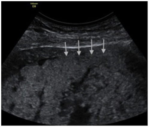
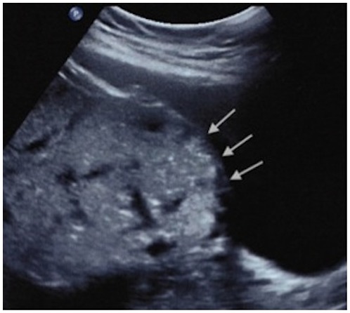
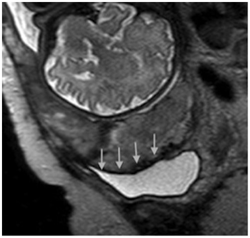
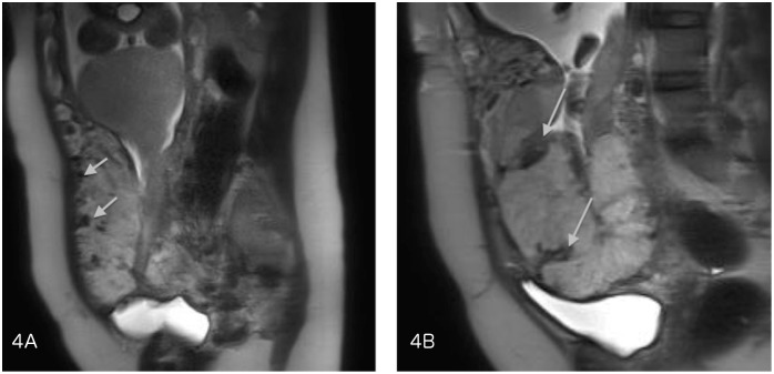
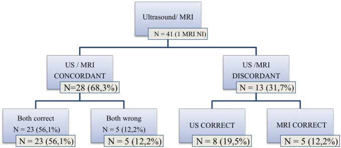
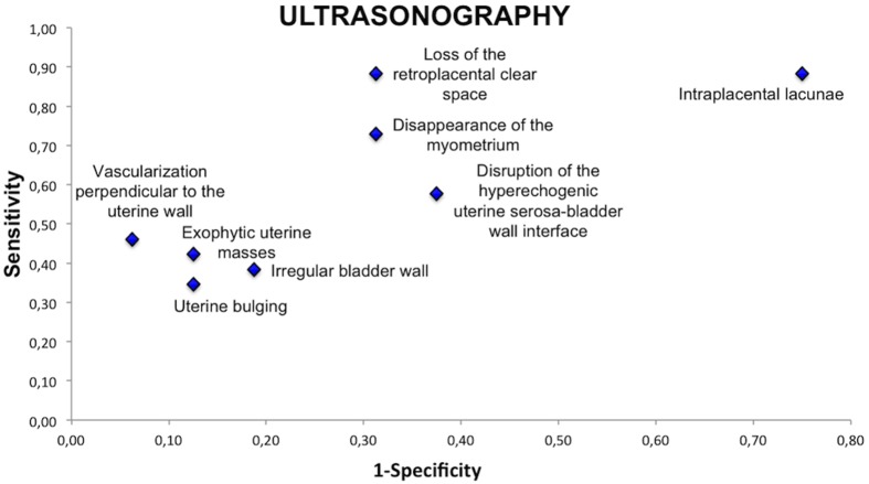
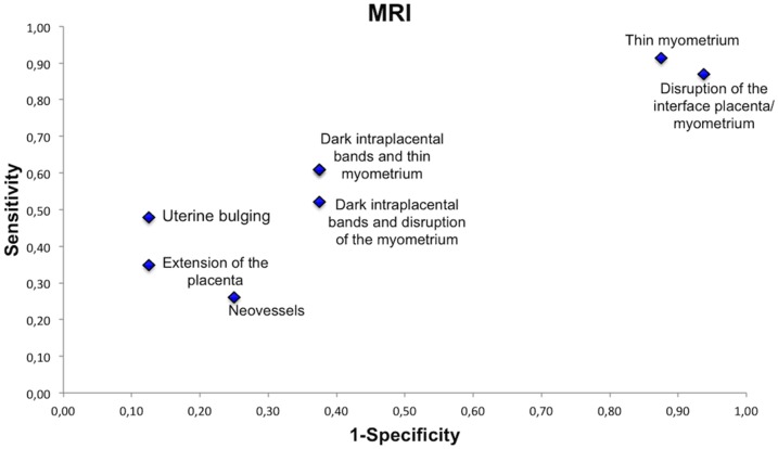
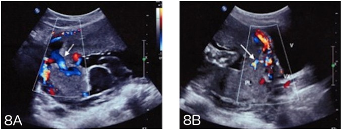
Results: Sensitivity in the diagnosis of placenta accreta was 100% with ultrasound and 76.9% for MRI (P = 0.03). Specificity was 37.5% with ultrasonography and 50% for MRI (P = 0.6). The features of greatest sensitivity on ultrasonography were intraplacental lacunae and loss of the normal retroplacental clear space. Increased vascularization in the uterine serosa-bladder wall interface and vascularization perpendicular to the uterine wall had the best positive predictive value (92%). At MRI, uterine bulging had the best positive predictive value (85%) and its combination with the presence of dark intraplacental bands on T2-weighted images improved the predictive value to 90%.
Conclusion: Ultrasound imaging is the mainstay of screening for placenta accreta. MRI appears to be complementary to ultrasonography, especially when there are few ultrasound signs.
Conflict of interest statement
Figures
References
MeSH terms
LinkOut - more resources
Full Text Sources
Other Literature Sources
Medical
