. 2015 Apr;73(4):1359-69.
doi: 10.1002/mrm.25256. Epub 2014 Apr 14.

Exchange kinetics by inversion transfer: integrated analysis of the phosphorus metabolite kinetic exchanges in resting human skeletal muscle at 7 T

Affiliations

Exchange kinetics by inversion transfer: integrated analysis of the phosphorus metabolite kinetic exchanges in resting human skeletal muscle at 7 T

Jimin Ren et al. Magn Reson Med. 2015 Apr.

Abstract

Purpose: To develop an inversion pulse-based, chemical exchange saturation transfer-like method for detection of (31) P magnetization exchanges among all nuclear magnetic resonance visible metabolites suitable for providing an integrated kinetic analysis of phosphorus exchange reactions in vivo.

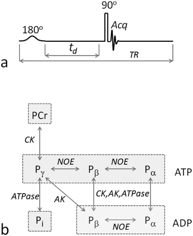
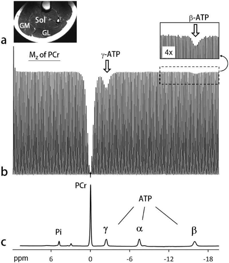
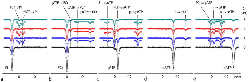
Methods: The exchange kinetics by inversion transfer (EKIT) sequence includes application of a frequency-selective inversion pulse arrayed over the range of relevant (31) P frequencies, followed by a constant delay and a hard readout pulse. A series of EKIT spectra, each given by a plot of Z-magnetization for each metabolite of interest versus frequency of the inversion pulse, can be generated from this single data set.

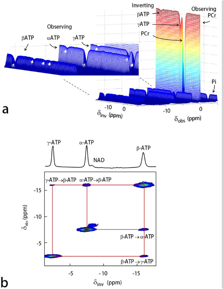
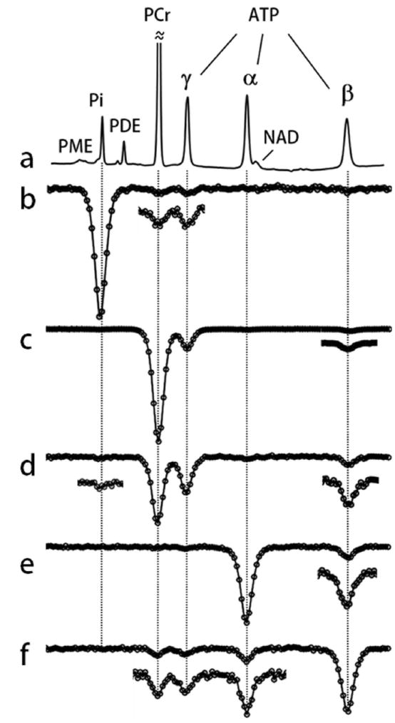
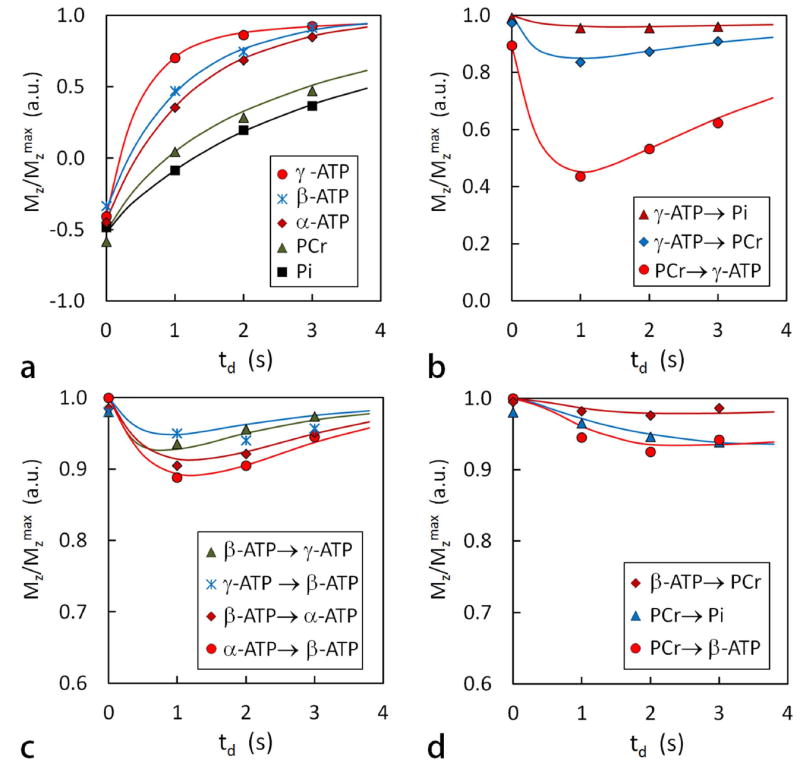
Results: EKIT spectra reflect chemical exchange due to known biochemical reactions, cross-relaxation effects, and relayed magnetization transfers due to both processes. The rate constants derived from EKIT data collected on resting human skeletal muscle were: ATP synthesis via ATP synthase (0.050 ± 0.016 s(-1) ), ATP synthesis via creatine kinase (0.264 ± 0.023 s(-1) ), and cross-relaxation between neighboring spin pairs within ATP (0.164 ± 0.022 s(-1) ).

Conclusion: EKIT provides a simple, alternative method to detect chemical exchange, cross relaxation, and relayed magnetization transfer effects in human skeletal muscle at 7 T.

Keywords: T1 relaxation time; chemical exchange; magnetization transfer; nuclear Overhauser effects; skeletal muscle.


Figures

FIG. 1
FIG. 1
EKIT pulse sequence and 31P magnetization exchange pathways. a: The EKIT pulse sequence consists of a low-power, frequency-sweep inversion pulse, followed by a constant delay period, td, and a hard readout pulse. b: 31P magnetization exchange system in skeletal muscle, showing the various exchange pathways that are possible in skeletal muscle, including phosphorous chemical exchange reactions indicated by vertical arrows, and Nuclear Overhauser Effect (NOE) indicated by horizontal arrows. ATP and ADP are marked by rectangular boxes. A five-pool model including Pi, PCr and ATP but not ADP, was adopted in this work. Abbreviations: AK, adenylate kinase; CK, creatine kinase.
FIG. 2
FIG. 2
EKIT spectrum of human calf skeletal muscle. a: A typical T2w MR image acquired from human calf using a partial volume coil. Abbreviations: Sol, soleus; GM, gastrocnemius medial; GL, gastrocnemius lateral. b: The EKIT spectrum illustrated here reflects the intensity of PCr signal plotted as a function of frequency of the inversion pulse (150 different frequencies) acquired from calf of a 25 yr healthy female with a BMI = 22.2 kg/m2. Other 31P MRS acquisition parameters: TR 5 second, td 1 second, scan time 12.5 min. Note that PCr magnetization is attenuated after inversion of either γ ATP (white arrow) or β ATP (insert). c: A conventional 31P MR spectrum acquired showing the full chemical shift region of calf muscle 31P resonances, with the chemical shift aligned with that of sweeping inversion pulse used in the EKIT spectrum b.
FIG. 3
FIG. 3
3D and 2D display of EKIT spectra of resting human calf muscle. a: 3D EKIT spectrum plotted by arraying the entire 31P MR spectra acquired using a series of offset inversion frequencies (PCr as reference at 0 ppm), with an enlarged region (inset) showing EKIT exchange peaks among the ATP spins (exchange effects marked by arrows with solid head, on-resonance inversion signals marked by arrows with thin lines). b: 2D contour plot showing ATP cross-relaxation (NOE) generated from subtraction spectra with off-resonance inversion as baseline. Note that the NAD peak which lies along the diagonal of this plot shows no exchange effect with ATP in resting skeletal muscle.
FIG. 4
FIG. 4
EKIT spectra of resting human calf muscle. a: A conventional 31P MR spectrum and (b-f) EKIT spectra with observation resonance at b: Pi, c: PCr, d: γ-ATP, e: α-ATP, and f: β-ATP acquired from resting human calf muscle at td = 1 s. These spectra represent averages from 7 subjects. In some spectra, small exchange peaks were enlarged 3-fold.
FIG. 5
FIG. 5
EKIT spectra and data fitting. EKIT spectra for observation of a: Pi, b: PCr, c: γ-ATP, d: α-ATP, and e: β-ATP at inversion delays of 0, 1, 2, and 3 seconds. The EKIT data were acquired from resting human calf muscle averaged for 7 subjects. The major chemical exchange reactions are indicated by solid black arrows while the effects of relayed MT are marked by the hollow arrows. The smaller EKIT exchange peaks were vertically enlarged (x4). The solid curves represent the least-squares fitting of all data to a five-pool kinetic exchange model shown in FIG. 1b.
FIG. 6
FIG. 6
Plots of normalized EKIT peak intensity versus delay time td for several exchange processes. a: plots of Mz/Mz max after an on-resonance inversion pulse for several metabolites, b: for chemical exchanges mediated by CK and ATPase, c: for NOE within ATP, and d: for relayed MT effects. These data are identical to those shown in FIG. 5 with the solid lines showing the best fit of the data to the exchange model.

References

    1. Brindle KM, Radda GK. Measurements of exchange in the reaction catalysed by creatine kinase using 14C and 15N isotope labels and the NMR technique of saturation transfer. Biochim Biophys Acta. 1985;829(2):188–201. - PubMed
    1. Brindle KM, Blackledge MJ, Challiss RA, Radda GK. 31P NMR magnetization-transfer measurements of ATP turnover during steady-state isometric muscle contraction in the rat hind limb in vivo. Biochemistry. 1989;28(11):4887–93. - PubMed
    1. Shoubridge EA, Briggs RW, Radda GK. 31p NMR saturation transfer measurements of the steady state rates of creatine kinase and ATP synthetase in the rat brain. FEBS Lett. 1982;140(2):289–92. - PubMed
    1. Lebon V, Dufour S, Petersen KF, Ren J, Jucker BM, Slezak LA, Cline GW, Rothman DL, Shulman GI. Effect of triiodothyronine on mitochondrial energy coupling in human skeletal muscle. J Clin Invest. 2001;108:733–737. - PMC - PubMed
    1. Schmid AI, Schrauwen-Hinderling VB, Andreas M, Wolzt M, Moser E, Roden M. Comparison of measuring energy metabolism by different (31) P-magnetic resonance spectroscopy techniques in resting, ischemic, and exercising muscle. Magn Reson Med. 2012;67(4):898–905. - PubMed

Publication types