. 2014 Jun;46(3):173-87.
doi: 10.1007/s10863-014-9549-9. Epub 2014 Apr 15.

MuRF1 activity is present in cardiac mitochondria and regulates reactive oxygen species production in vivo

Affiliations

MuRF1 activity is present in cardiac mitochondria and regulates reactive oxygen species production in vivo

Taylor A Mattox et al. J Bioenerg Biomembr. 2014 Jun.

Erratum in

Abstract

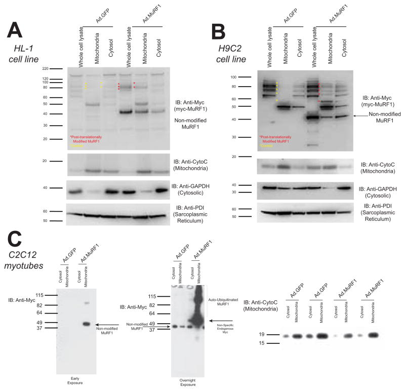
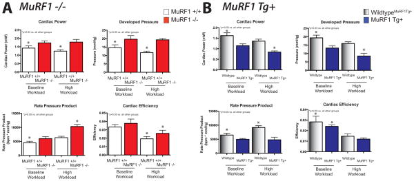
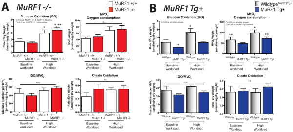
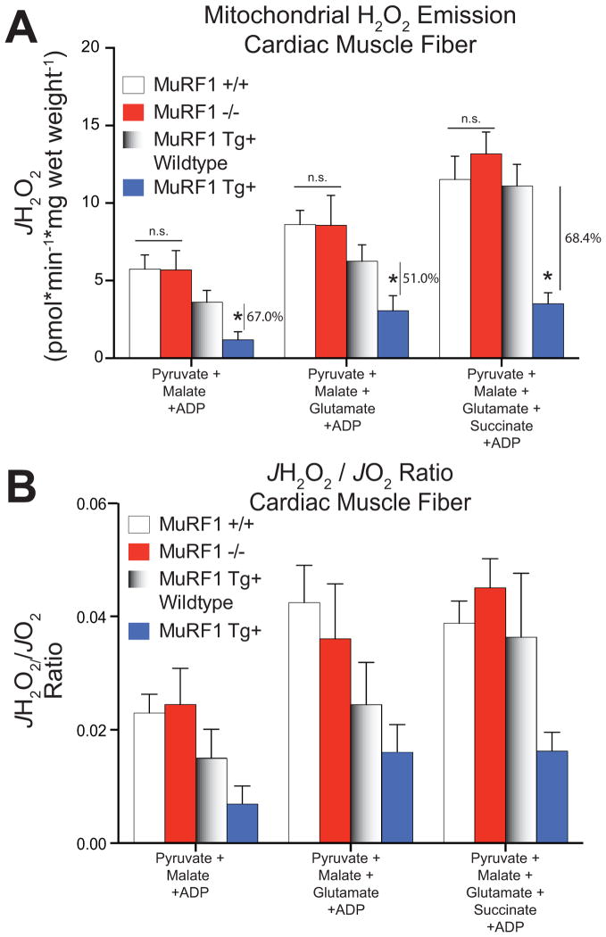
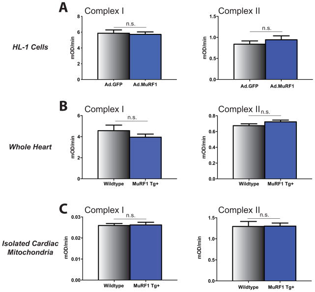
MuRF1 is a previously reported ubiquitin-ligase found in striated muscle that targets troponin I and myosin heavy chain for degradation. While MuRF1 has been reported to interact with mitochondrial substrates in yeast two-hybrid studies, no studies have identified MuRF1's role in regulating mitochondrial function to date. In the present study, we measured cardiac mitochondrial function from isolated permeabilized muscle fibers in previously phenotyped MuRF1 transgenic and MuRF1-/- mouse models to determine the role of MuRF1 in intermediate energy metabolism and ROS production. We identified a significant decrease in reactive oxygen species production in cardiac muscle fibers from MuRF1 transgenic mice with increased α-MHC driven MuRF1 expression. Increased MuRF1 expression in ex vivo and in vitro experiments revealed no alterations in the respiratory chain complex I and II function. Working perfusion experiments on MuRF1 transgenic hearts demonstrated significant changes in glucose oxidation. However, total oxygen consumption was decreased [corrected]. This data provides evidence for MuRF1 as a novel regulator of cardiac ROS, offering another mechanism by which increased MuRF1 expression may be cardioprotective in ischemia reperfusion injury, in addition to its inhibition of apoptosis via proteasome-mediate degradation of c-Jun. The lack of mitochondrial function phenotype identified in MuRF1-/- hearts may be due to the overlapping interactions of MuRF1 and MuRF2 with energy regulating proteins found by yeast two-hybrid studies reported here, implying a duplicity in MuRF1 and MuRF2's regulation of mitochondrial function.


Conflict of interest statement

Disclosures

There are no conflicts of interest to disclose.

Figures

Figure 1
Figure 1. MuRF1 is found present in the mitochondria
Western analysis of MuRF1 expression in mitochondrial and cytosolic fractions in HL-1 and H9C2 cells. Cells were transduced with Ad.mycMuRF1 or Ad.GFP and lysed to obtain whole cell lysate or differential centrifugation to isolate mitochondria and cytosol fractions. Immunoblot for A. MuRF1 (anti-myc), cytochrome c, GAPDH, and PDI illustrate MuRF1’s presence in the mitochondria and auto-ubiquitination. These unique auto-ubiquitinated forms of (myc)MuRF1 can be seen in the mitochondria and cytosol fractions in both the HL-1 atrial cardiomyocyte derived, B. H9C2 ventricular cardiomyocyte derived cell line, as well as the C. C2C12 undifferentiated myotubes. MuRF1 (anti-myc) and cytochrome c (mitochondria), illustrated at both early (left, 30 second exposure) and extended (right-overnight) exposures.
Figure 2
Figure 2. MuRF1−/− hearts exhibit increase power and developed pressure at baseline and high workload conditions while MuRF1 Tg+ hearts have impaired
Functional characteristics of A. MuRF1−/− and B. MuRF1 Tg+ heart function ex vivo. Hearts were isolated and immediate perfused in the working mode at basal (50 mm Hg) and high (80 mm Hg) workloads. Cardiac power (a derivative of cardiac output), cardiac efficiency (ratio of cardiac work and myocardial oxygen consumption), and rate pressure product (heart rate*developed pressure) were calculated as previously described [21]. Values are expressed as mean ± SEM and represent 5–7 mice per group. Groups were analyzed by a One Way ANOVA vs. sibling match wildtype controls at baseline and high workload conditions. n.s.= not significant. p<0.05, unless otherwise noted in the panel.
Figure 3
Figure 3. MuRF1 Tg+ hearts exhibits alterations in glucose oxidation and oxygen consumption ex vivo
Metabolic (glucose and oleate oxidation) and oxygen consumption utilization of A. MuRF1−/− and B. MuRF1 Tg+ hearts compared to strain matched wildtype controls. Hearts were isolated and immediate perfused in the working mode at basal (50 mm Hg) and high (80 mm Hg) workloads. Values are expressed as mean ± SEM and represent 5–7 mice per group. Groups were analyzed by a One Way ANOVA vs. sibling match wildtype controls at baseline and high workload conditions. n.s.= not significant. MVO2=myocardial oxygen consumption. p<0.05, unless otherwise noted in the panel.
Figure 4
Figure 4. Increasing MuRF1 expression inhibits Krebs cycle flux in permeabilized cardiac muscle fibers
A. Quantified rates of pyruvate/malate-supported O2 consumption (JO2) in permeabilized left ventricle myofibers from MuRF Tg+, MuRF −/−, and their respective WT controls. B. No differences in mitochondrial coupling were detected as indicated by RCR, represents the ratio of state 3 (phosphorylating) respiration to state 4 respiration. C. Quantified rates of palmitoyl-L-carnitine -supported O2 consumption in permeabilized left ventricle myofibers from MuRF Tg+, MuRF −/−, and their respective WT controls. D. Quantification of calcium retention capacity in isolated cardiac mitochondria from MuRF Tg+, MuRF −/−, and their respective WT controls. Data are expressed as mean ± SD and are representative of 3–5 mice per group. *p<0.05 by Student’s t-test vs. sibling-matched wildtype controls.
Figure 5
Figure 5. Mitochondrial H2O2 emission is inhibited in permeabilized cardiac muscle fibers from MuRF1 Tg+ hearts
A. Quantified rates of mH2O2 emission (JH2O2) and B. the JH2O2/JO2 ratio from permeabilized muscle fibers obtained from MuRF Tg+, MuRF−/−, and their respective controls. Data are expressed as mean ± SD and are representative of 3–5 mice per group. *p<0.05 by Student’s t-test vs. sibling-matched wildtype controls.
Figure 6
Figure 6. Mitochondrial Complex I/Complex II Activity with increased MuRF1 expression in HL-1 cells, MuRF1 Tg+ cardiac ventricular lysates and MuRF1 Tg+ isolated cardiac mitochondria
Microplate activity assays measuring mitochondrial complex I activity (oxidation of NADH to NAD+) and complex II activity (reduction of ubiquinone to ubiquinol) in MuRF1 overexpressing HL-1 cells, MuRF1 Tg+ cardiac ventricular lysates and isolated cardiac mitochondria from MuRF1 Tg+ mice demonstrate no significant difference in complex I or complex II activity as a consequence of MuRF1 overexpression. (a) Mitochondrial complex I and complex II activity in total protein lysates isolated from HL-1 cells transduced with adenovirus expressing CMV GFP control or CMV GFP-tagged myc-MURF1. (b) Mitochondrial complex I and complex II activity in whole cardiac ventricular lysates from WT and MuRF1 Tg+ mice. (c) Mitochondrial complex I and complex II activity in mitochondria isolated from WT and MuRF1 Tg+ mouse hearts. n.s.= not significant by Student’s t-test vs. sibling-matched wildtype controls.

Similar articles

Cited by

References

    1. Bodine SC, Latres E, Baumhueter S, Lai VK, Nunez L, Clarke BA, et al. Identification of ubiquitin ligases required for skeletal muscle atrophy. Science. 2001;294(5547):1704–1708. doi: 10.1126/science.1065874. 1065874 [pii] - DOI - PubMed
    1. Willis MS, Rojas M, Li L, Selzman CH, Tang RH, Stansfield WE, et al. Muscle ring finger 1 mediates cardiac atrophy in vivo. Am J Physiol Heart Circ Physiol. 2009;296(4):H997–H1006. doi: 10.1152/ajpheart.00660.2008. 00660.2008 [pii] - DOI - PMC - PubMed
    1. Centner T, Yano J, Kimura E, McElhinny AS, Pelin K, Witt CC, et al. Identification of muscle specific ring finger proteins as potential regulators of the titin kinase domain. [Research Support, Non-U.S. Gov’t Research Support, U.S. Gov’t, P.H.S.] J Mol Biol. 2001;306(4):717–726. doi: 10.1006/jmbi.2001.4448. - DOI - PubMed
    1. Arya R, Kedar V, Hwang JR, McDonough H, Li HH, Taylor J, et al. Muscle ring finger protein-1 inhibits PKC{epsilon} activation and prevents cardiomyocyte hypertrophy. J Cell Biol. 2004;167(6):1147–1159. doi: 10.1083/jcb.200402033. jcb.200402033 [pii] - DOI - PMC - PubMed
    1. Witt SH, Granzier H, Witt CC, Labeit S. MURF-1 and MURF-2 target a specific subset of myofibrillar proteins redundantly: towards understanding MURF-dependent muscle ubiquitination. J Mol Biol. 2005;350(4):713–722. doi: 10.1016/j.jmb.2005.05.021. S0022-2836(05)00552-8 [pii] - DOI - PubMed

Publication types

LinkOut - more resources