. 2014 Apr 29;111(17):6305-10.
doi: 10.1073/pnas.1321406111. Epub 2014 Apr 14.

X-ray structure of human aquaporin 2 and its implications for nephrogenic diabetes insipidus and trafficking

Affiliations

X-ray structure of human aquaporin 2 and its implications for nephrogenic diabetes insipidus and trafficking

Anna Frick et al. Proc Natl Acad Sci U S A. .

Abstract

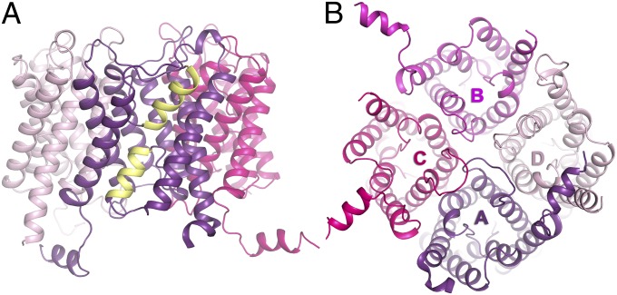
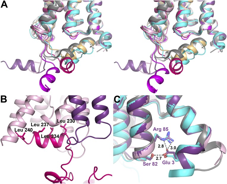
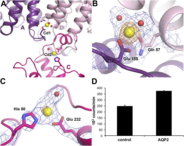
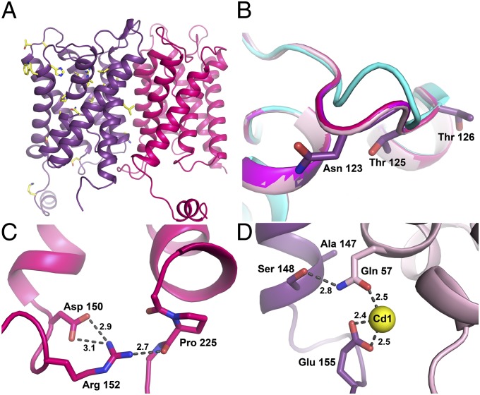
Human aquaporin 2 (AQP2) is a water channel found in the kidney collecting duct, where it plays a key role in concentrating urine. Water reabsorption is regulated by AQP2 trafficking between intracellular storage vesicles and the apical membrane. This process is tightly controlled by the pituitary hormone arginine vasopressin and defective trafficking results in nephrogenic diabetes insipidus (NDI). Here we present the X-ray structure of human AQP2 at 2.75 Å resolution. The C terminus of AQP2 displays multiple conformations with the C-terminal α-helix of one protomer interacting with the cytoplasmic surface of a symmetry-related AQP2 molecule, suggesting potential protein-protein interactions involved in cellular sorting of AQP2. Two Cd(2+)-ion binding sites are observed within the AQP2 tetramer, inducing a rearrangement of loop D, which facilitates this interaction. The locations of several NDI-causing mutations can be observed in the AQP2 structure, primarily situated within transmembrane domains and the majority of which cause misfolding and ER retention. These observations provide a framework for understanding why mutations in AQP2 cause NDI as well as structural insights into AQP2 interactions that may govern its trafficking.

Keywords: X-ray crystallography; membrane protein; water channel protein.


Conflict of interest statement

The authors declare no conflict of interest.

Figures

Fig. 1.
Fig. 1.
Overall structure of human AQP2. (A) Overview of the AQP2 tetramer with half helices formed by loops B and E highlighted in yellow. (B) Overview of the AQP2 tetramer from the intracellular side. The color scheme for each of the protomers is used throughout the article (A, purple; B, magenta; C, pink; D, light pink).
Fig. 2.
Fig. 2.
Structure of the C and N termini. (A) Stereo image of the four AQP2 protomers (colored as in Fig. 1) overlaid on OaAQP0 [white; Protein Data Bank (PDB) ID code 2B60], BtAQP0 (light orange; PDB ID code 1YMG), BtAQP1 (gray; PDB ID code 1J4N), and HsAQP5 (cyan; PDB ID code 3D9S). The C termini of all four AQP2 protomers occupy different positions, none of which overlay with any of the previous AQP structures. (B) The crystal contact site between the C-terminal helix of monomer C (pink) and a symmetry-related protomer D (light pink). Leucines lining up on one side of the helix are labeled. (C) Overlay of the N termini of protomer A and D with HsAQP5 (cyan) and BtAQP1 (gray). For protomer A (purple), Glu3 interacts with Ser82 and Arg85, similar to the structural arrangement seen in AQP5. In contrast, protomer D (light pink) resembles BtAQP1 with TM helix 1 extending a full turn into the cytoplasm.
Fig. 3.
Fig. 3.
Cd2+ binding sites in AQP2. (A) Two Cd2+ binding sites (Cd1 and Cd2) are found on the cytoplasmic side of the AQP2 tetramer. Yellow spheres represent Cd2+. The interaction between the C-terminal helix of protomer C (pink) with protomer D (light pink) of a symmetry-related tetramer is also shown. (B) Electron density for the Cd1 site showing 2Fobs–Fcalc map contoured at 1.5 σ (blue) and anomalous difference map contoured at 3.5 σ (orange). Residues serving as Cd2+ ligands are shown in stick representation. Water molecules ligating the Cd2+ ion are shown as red spheres. (C) Electron density for the Cd2 site showing a 2Fobs–Fcalc map contoured at 1.2 σ (blue) and anomalous difference map contoured at 3.5 σ (orange). Cd2+ ligands are displayed as in B. (D) Radioactive Ca2+ binding assay illustrating how AQP2-expressing Xenopus oocytes bind significantly more 45Ca2+ (P < 0.05) than controls, suggesting that Ca2+ is the physiological ligand for the Cd+ binding sites. Mean ± SEM is given of three groups of 10 oocytes per condition.
Fig. 4.
Fig. 4.
NDI-causing mutations in AQP2. (A) Overview of known NDI-causing mutations (yellow) in the AQP2 structure showing how these mostly affect the pore-forming regions. (B) Structural overlay of loop C from the four AQP2s showing the N-glycosylation site at Asn123. A comparison with other mammalian AQPs, here represented by AQP5 (cyan), shows that structure of AQP2 is markedly different at this site. Asn123 as well as two nearby NDI mutation sites, Thr125 and Thr126, are shown in stick representation. (C) Structure around the NDI mutation site at Asp150, showing its connections to Pro225 at the proximal end of the C-terminal tail via Arg152. Hydrogen bonds are indicated by dotted lines. Distances are shown in Å. (D) Close-up of the Cd1 site showing how Ser148 hydrogen bonds to the Cd2+ ligand Gln57. Mutation of both these residues cause ER retention of AQP2. Bonds are indicated by dotted lines with distances shown in Å.

Similar articles

Cited by

References

    1. Verbalis JG. Disorders of body water homeostasis. Best Pract Res Clin Endocrinol Metab. 2003;17(4):471–503. - PubMed
    1. van Os CH, Deen PM, Dempster JA. Aquaporins: Water selective channels in biological membranes. Molecular structure and tissue distribution. Biochim Biophys Acta. 1994;1197(3):291–309. - PubMed
    1. Noda Y, Sohara E, Ohta E, Sasaki S. Aquaporins in kidney pathophysiology. Nat Rev Nephrol. 2010;6(3):168–178. - PubMed
    1. Fushimi K, Sasaki S, Marumo F. Phosphorylation of serine 256 is required for cAMP-dependent regulatory exocytosis of the aquaporin-2 water channel. J Biol Chem. 1997;272(23):14800–14804. - PubMed
    1. Katsura T, Gustafson CE, Ausiello DA, Brown D. Protein kinase A phosphorylation is involved in regulated exocytosis of aquaporin-2 in transfected LLC-PK1 cells. Am J Physiol. 1997;272(6 Pt 2):F817–F822. - PubMed

Publication types

Associated data