. 2014 Apr 29;111(17):6509-14.
doi: 10.1073/pnas.1402243111. Epub 2014 Apr 14.

Structural basis for regulation of rhizobial nodulation and symbiosis gene expression by the regulatory protein NolR

Affiliations

Structural basis for regulation of rhizobial nodulation and symbiosis gene expression by the regulatory protein NolR

Soon Goo Lee et al. Proc Natl Acad Sci U S A. .

Abstract

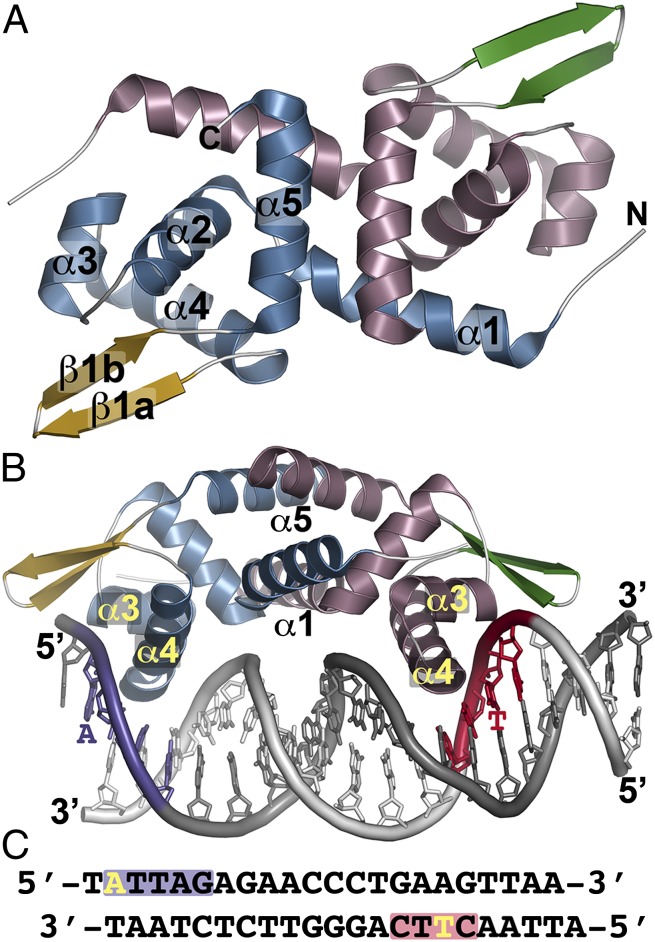
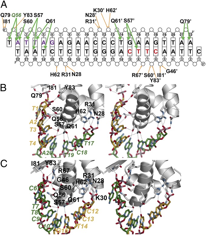
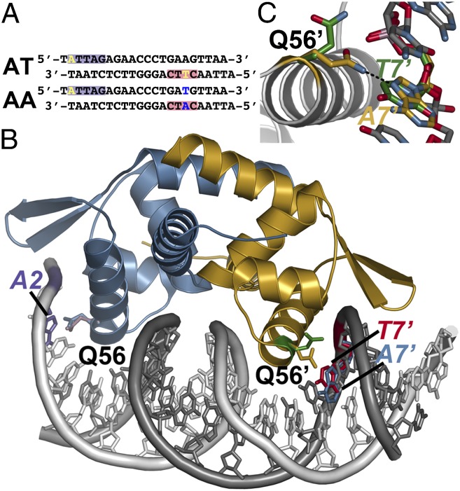
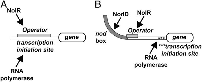
The symbiosis between rhizobial microbes and host plants involves the coordinated expression of multiple genes, which leads to nodule formation and nitrogen fixation. As part of the transcriptional machinery for nodulation and symbiosis across a range of Rhizobium, NolR serves as a global regulatory protein. Here, we present the X-ray crystal structures of NolR in the unliganded form and complexed with two different 22-base pair (bp) double-stranded operator sequences (oligos AT and AA). Structural and biochemical analysis of NolR reveals protein-DNA interactions with an asymmetric operator site and defines a mechanism for conformational switching of a key residue (Gln56) to accommodate variation in target DNA sequences from diverse rhizobial genes for nodulation and symbiosis. This conformational switching alters the energetic contributions to DNA binding without changes in affinity for the target sequence. Two possible models for the role of NolR in the regulation of different nodulation and symbiosis genes are proposed. To our knowledge, these studies provide the first structural insight on the regulation of genes involved in the agriculturally and ecologically important symbiosis of microbes and plants that leads to nodule formation and nitrogen fixation.

Keywords: protein structure; transcription factor.


Conflict of interest statement

The authors declare no conflict of interest.

Figures

Fig. 1.
Fig. 1.
Overall structure of NolR. (A) The structure of unliganded NolR is shown as a ribbon diagram. Secondary structure features are labeled in monomer A and are differentially colored in each monomer as follows: blue α-helices and gold β-strands in monomer A and rose α-helices and green β-strands in monomer b. This is a “top” view of the dimeric structure. (B) Structure of NolR in complex with the 22-bp oligo AT duplex. Secondary structure features are colored as in A. The view is rotated ∼90° relative to A to present a “side” view of interaction with DNA. α-Helices forming the dimer interface (α1 and α5) and the helix-turn-helix motif (α3–α4) are labeled. Consensus-motif regions of oligo AT that contact NolR are colored purple and red with key nucleotides indicated. (C) Sequence of oligo AT. The purple and red boxes correspond to the regions of the consensus motif highlighted in B. The yellow A and T indicate nucleotides that are variable in the target DNA sequences of NolR.
Fig. 2.
Fig. 2.
NolR and asymmetric operator binding. (A) Schematic showing NolR-oligo AT DNA contacts. The bases are labeled and shown as rectangles, with phosphate and ribose groups drawn as circles and pentagons, respectively. Residues from chain B of the homodimer are noted with an apostrophe after the amino acid number. Orange arrows indicate backbone contacts, and green arrows show base-specific interactions. The two halves of the NolR consensus target sequence that interact with NolR are highlighted with purple and red color, as in Fig. 1 B and C. Gln56 is colored green to emphasize its role in consensus-motif recognition. (B) Stereoview of protein–DNA interactions in the first half-site. Protein side-chains are from chain A. Nucleotides from 5′ and 3′ strands are colored gold and green, respectively, and are labeled. (C) Stereoview of protein–DNA interactions in the second half-site. Protein side-chains are from chain B. Nucleotides from 5′ and 3′ strands are colored gold and green, respectively, and are labeled.
Fig. 3.
Fig. 3.
Conformational switching of Gln56 for recognition of variable DNA target sites by NolR. (A) Sequences of oligos AT and AA. The purple and red boxes correspond to the regions of the consensus motif highlighted in Fig. 1 B and C. The variable position bases are highlighted yellow in oligo AT. Changes at these positions in oligo AA are highlighted in blue. (B) Structure of NolR in complex with the 22-bp oligo AA duplex. Chains A and B of NolR are colored blue and gold, respectively. The 5′ and 3′ strands of oligo AA are colored white and gray, respectively. The orientation of the Gln56 side-chain in each monomer of NolR complexed with either oligo AT (purple and green sticks) or oligo AA (blue and gold sticks) is shown. The positions of A2 (purple) from oligo AA, T7' (red) from oligo AT, and A7' (blue) from oligo AA are shown. (C) Close-up view of Gln56 movement in the second consensus half-site of NolR. The position of Gln56 of chain B and the variable base is shown. The structures observed with NolR complexed with either oligo AT (green) and oligo AA (gold) are shown.
Fig. 4.
Fig. 4.
Models of NolR regulation of nodulation and symbiosis gene expression. (A) In promoters with overlapping transcription initiation and operator sites, NolR binding prevents RNA polymerase interaction and gene expression. (B) In promoter regions containing upstream nod box sequences for binding of the transcriptional activator NodD, binding of NolR to the operator site may either alter association of NodD to the nod box or alter DNA bending that results from NodD binding to prevent activation of gene expression.

Similar articles

Cited by

References

    1. Desbrosses GJ, Stougaard J. Root nodulation: A paradigm for how plant-microbe symbiosis influences host developmental pathways. Cell Host Microbe. 2011;10(4):348–358. - PubMed
    1. Kondorosi E, Mergaert P, Kereszt A. A paradigm for endosymbiotic life: Cell differentiation of Rhizobium bacteria provoked by host plant factors. Annu Rev Microbiol. 2013;67:611–628. - PubMed
    1. Masson-Boivin C, Giraud E, Perret X, Batut J. Establishing nitrogen-fixing symbiosis with legumes: How many rhizobium recipes? Trends Microbiol. 2009;17(10):458–466. - PubMed
    1. Horvath B, et al. Organization, structure and symbiotic function of Rhizobium meliloti nodulation genes determining host specificity for alfalfa. Cell. 1986;46(3):335–343. - PubMed
    1. Long SR. Rhizobium symbiosis: Nod factors in perspective. Plant Cell. 1996;8(10):1885–1898. - PMC - PubMed

MeSH terms

LinkOut - more resources