. 2014 Apr 29;111(17):6425-30.
doi: 10.1073/pnas.1321507111. Epub 2014 Apr 14.

Tetherin antagonism by Vpu protects HIV-infected cells from antibody-dependent cell-mediated cytotoxicity

Affiliations

Tetherin antagonism by Vpu protects HIV-infected cells from antibody-dependent cell-mediated cytotoxicity

Juan F Arias et al. Proc Natl Acad Sci U S A. .

Abstract

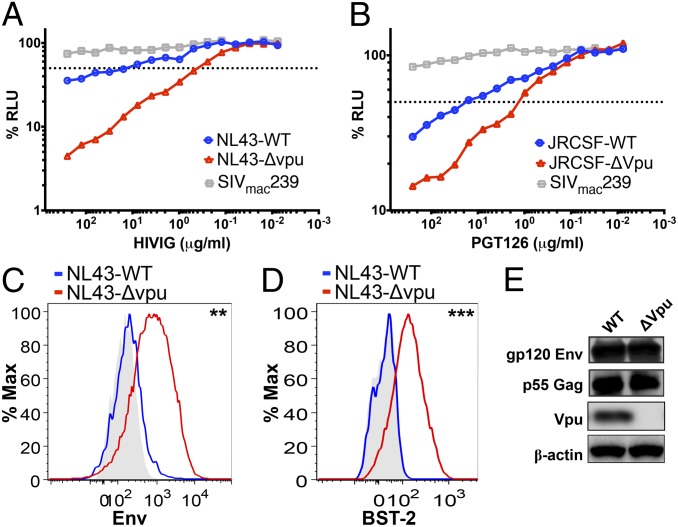
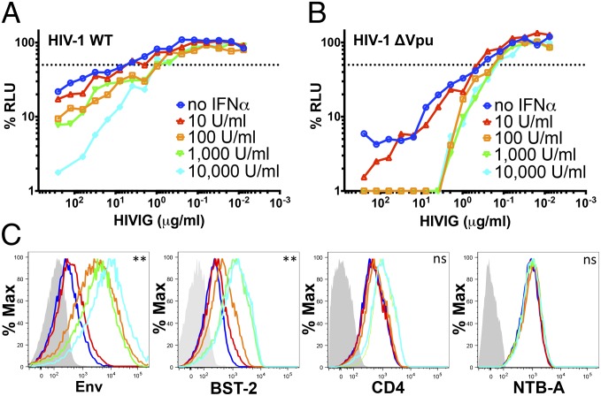
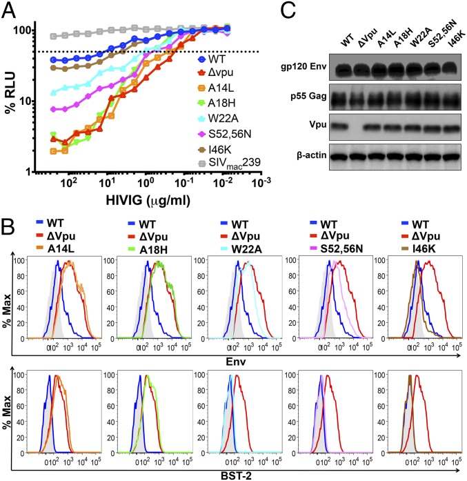
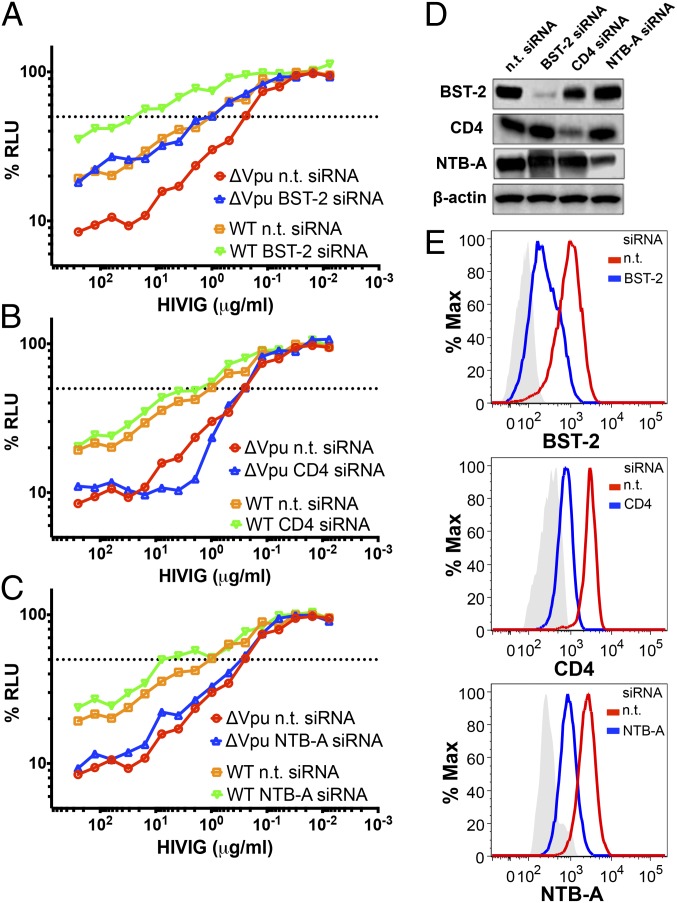
Tetherin is an IFN-inducible transmembrane protein that inhibits the detachment of enveloped viruses from infected cells. HIV-1 overcomes this restriction factor by expressing HIV-1 viral protein U (Vpu), which down-regulates and degrades tetherin. We report that mutations in Vpu that impair tetherin antagonism increase the susceptibility of HIV-infected cells to antibody-dependent cell-mediated cytotoxicity (ADCC), and conversely that RNAi knockdown of tetherin, but not other cellular proteins down-modulated by Vpu, decreases the susceptibility of HIV-infected cells to ADCC. These results reveal that Vpu protects HIV-infected cells from ADCC as a function of its ability to counteract tetherin. By serving as link between innate and adaptive immunity, the antiviral activity of tetherin may be augmented by virus-specific antibodies, and hence much greater than previously appreciated.

Keywords: AIDS; BST-2; CD317; lentivirus.


Conflict of interest statement

The authors declare no conflict of interest.

Figures

Fig. 1.
Fig. 1.
Deletion of vpu increases the susceptibility of HIV-infected cells to ADCC. CEM.NKR-CCR5-sLTR-Luc target cells containing a Tat-inducible luciferase reporter gene were infected with WT vs. vpu-deleted HIV-1NL4-3 (A) or HIV-1JR-CSF (B) and incubated at a 10:1 effector:target ratio with an NK cell line that constitutively expresses CD16 in the presence of serial dilutions of antibody, either purified Ig from HIV-1+ donors (i.e., HIVIG) or the gp120-specific monoclonal antibody PGT126. SIVmac239-infected target cells were included as a control for nonspecific killing. ADCC activity was measured as the dose-dependent loss of luciferase activity in RLU over an 8-h incubation as previously described (21). The dotted line indicates 50% killing of HIV-infected cells. Surface expression of HIV-1 Env (C) and tetherin (D) was compared on cells infected with WT vs. vpu-deleted HIV-1. CEM.NKR-CCR5-sLTR-Luc cells infected with WT HIV-1 (blue lines) and vpu-deleted HIV-1 (red lines) were stained for Env and tetherin (BST-2), followed by permeabilization and intracellular staining for Gag as described in Materials and Methods. Histograms represent the fluorescence intensity of surface staining for Env (C) and tetherin (D) after gating on viable, virus-infected (Gag+) CD45+ cells relative to control samples (shaded) stained with normal human IgG (C) or an isotype control for the BST-2–specific antibody (D). Differences in the geometric mean fluorescence intensity were significant for Env (**P = 0.01, unpaired t test) (C) and tetherin (D; ***P = 0.00009). (E) Immunoblot analysis of total levels of Env, Gag, and Vpu expression in whole-cell lysates of 293T cells (tetherin-negative) transfected with WT or vpu-deleted HIV-1NL4-3 proviral DNA. The membrane was subsequently reprobed with a β-actin–specific antibody to control for sample loading. The data in AE are representative of three different experiments. A comparison of the data from three separate ADCC assays is provided in Fig. S1.
Fig. 2.
Fig. 2.
Treatment with IFN-α enhances the susceptibility of HIV-infected cells to ADCC. HIV-1NL4-3 (A) and HIV-1NL4-3 Δvpu (B)–infected CEM.NKR-CCR5-sLTR-Luc cells were treated with the indicated concentrations of IFN-α 24 h before they were used as target cells in an ADCC assay with serial dilutions of HIVIG. The dotted line indicates 50% killing of HIV-infected cells. A comparison of the data from three separate ADCC assays is provided in Fig. S2. (C) Changes in the surface expression of Env, tetherin (BST-2), CD4, and NTB-A on HIV-1 Δvpu-infected cells in response to IFN-α were measured by flow cytometry as described in Materials and Methods. The colored lines correspond to the indicated concentrations of IFN-α in A, and the shaded histograms indicate background staining with normal human IgG or an isotype control antibody. Differences in the geometric mean fluorescence intensity of staining were significant for cells treated with 0 vs. 10,000 U/mL IFN-α for Env (**P = 0.004, unpaired t test) and for tetherin (**P = 0.002), but not for CD4 or NTB-A (not significant). ADCC responses and surface expression of tetherin, but not CD4 or NTB-A, correlated with surface levels of Env (Fig. S2 GJ).
Fig. 3.
Fig. 3.
Substitutions in Vpu that impair tetherin antagonism increase the sensitivity of HIV-infected cells to ADCC. (A) CEM.NKR-CCR5-sLTR-Luc cells infected with HIV-1NL4-3 mutants with Vpu substitutions A14L, A18H, W22A, S52,56N, and I46K were tested for susceptibility to ADCC in the presence of the indicated concentrations of HIVIG. Target cells infected with WT HIV-1NL4-3, HIV-1NL4-3 Δvpu, and SIVmac239 were also included as controls. The dotted line indicates 50% ADCC activity. A comparison of data from three independent experiments is provided in Fig. S3 AC. (B) Surface expression of Env and tetherin (BST-2) for CEM.NKR-CCR5-sLTR-Luc cells infected with each Vpu mutant was compared with cells infected with WT (blue lines) and vpu-deleted HIV-1NL4-3 (red lines). The color scheme for each of the mutants is the same as in A. The histogram plots represent the fluorescence intensity of surface staining for Env (Upper) and tetherin (Lower) in viable virus-infected (Gag+) CD45+ cells in relation to control samples (shaded) stained with normal human IgG (Upper) or with an isotype control for the BST-2-specific antibody (Lower). ADCC responses and surface expression of tetherin, but not CD4 or NTB-A, correlated with surface levels of Env (Fig. S3 DG). (C) Immunoblot analysis of total levels of Env, Gag, and Vpu expression in whole-cell lysates of 293T cells (tetherin-negative) transfected with proviral DNA for WT HIV-1NL4-3 or each of the indicated vpu mutants. Reprobing with a β-actin–specific antibody was used to control for sample loading.
Fig. 4.
Fig. 4.
RNAi knockdown of tetherin increases the resistance of HIV-infected cells to ADCC. CEM.NKR-CCR5-sLTR-Luc cells infected with WT and vpu-deleted HIV-1NL4-3 were treated with nontargeting (n.t.) siRNA and siRNAs specific for tetherin (A), CD4 (B), and NTB-A (C) 48 h before they were used as target cells in an ADCC assay with HIVIG. The dotted line indicates 50% killing of HIV-infected cells. RNAi knockdown of tetherin (BST-2), CD4, and NTB-A was confirmed by immunoblot analysis of cell lysates (D) and by flow cytometry (E). (D) Immunoblot analysis of tetherin, CD4, and NTB-A expression in whole-cell lysates of HIV-1 Δvpu-infected CEM.NKR-CCR5-sLTR-Luc cells treated with a nontargeting (n.t.) siRNA or specific siRNAs targeting tetherin, CD4, or NTB-A. Reprobing with a β-actin–specific antibody was used to control for sample loading. Histogram plots in E represent the change in surface expression of tetherin, CD4, and NTB-A on HIV-1 Δvpu-infected target cells treated with specific siRNAs (blue lines) targeting tetherin (Top), CD4 (Middle), or NTB-A (Bottom) in relation to samples treated with a nontargeting (n.t.) siRNA (red lines). The shaded histograms indicate background staining with an isotype control antibody. A comparison of data from three independent experiments is provided in Fig. S4.

References

    1. Perez-Caballero D, et al. Tetherin inhibits HIV-1 release by directly tethering virions to cells. Cell. 2009;139(3):499–511. - PMC - PubMed
    1. Hammonds J, Wang J-J, Yi H, Spearman P. Immunoelectron microscopic evidence for Tetherin/BST2 as the physical bridge between HIV-1 virions and the plasma membrane. PLoS Pathog. 2010;6(2):e1000749. - PMC - PubMed
    1. Fitzpatrick K, et al. Direct restriction of virus release and incorporation of the interferon-induced protein BST-2 into HIV-1 particles. PLoS Pathog. 2010;6(3):e1000701. - PMC - PubMed
    1. Kupzig S, et al. Bst-2/HM1.24 is a raft-associated apical membrane protein with an unusual topology. Traffic. 2003;4(10):694–709. - PubMed
    1. VanDamme N, et al. The interferon-induced protein BST-2 restricts HIV-1 release and is downregulated from the cell surface by the viral vpu protein. Cell Host Microbe. 2008;3(4):245–252. - PMC - PubMed

Publication types

MeSH terms

Substances

LinkOut - more resources