

Reactive cysteine persulfides and S-polythiolation regulate oxidative stress and redox signaling
- PMID: 24733942
- PMCID: PMC4040604
- DOI: 10.1073/pnas.1321232111
Reactive cysteine persulfides and S-polythiolation regulate oxidative stress and redox signaling
Abstract
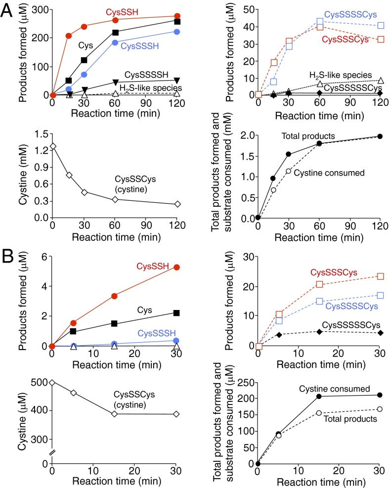
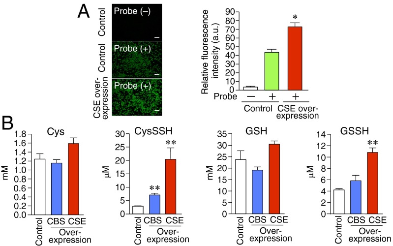
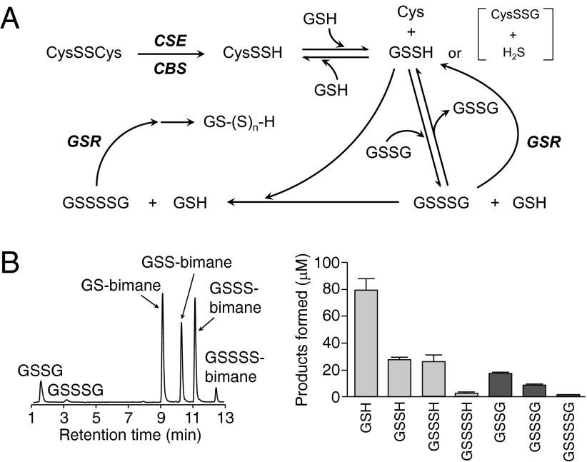
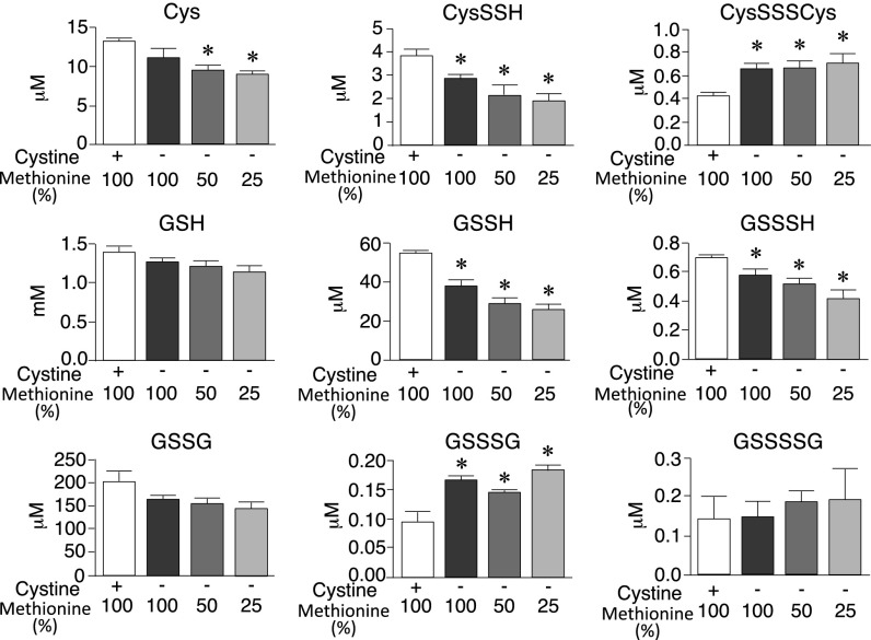
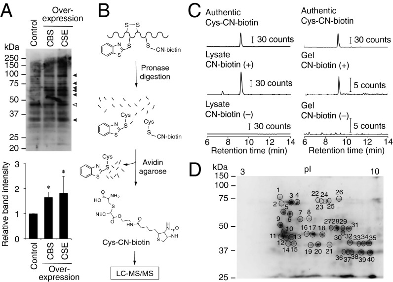
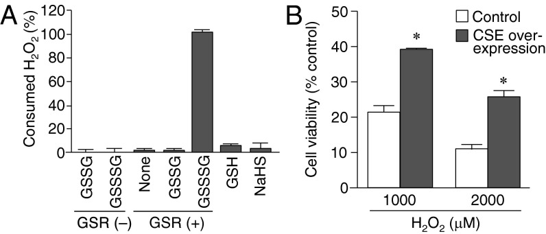
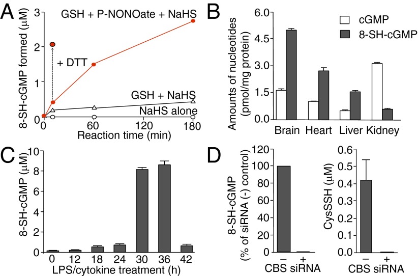
Using methodology developed herein, it is found that reactive persulfides and polysulfides are formed endogenously from both small molecule species and proteins in high amounts in mammalian cells and tissues. These reactive sulfur species were biosynthesized by two major sulfurtransferases: cystathionine β-synthase and cystathionine γ-lyase. Quantitation of these species indicates that high concentrations of glutathione persulfide (perhydropersulfide >100 μM) and other cysteine persulfide and polysulfide derivatives in peptides/proteins were endogenously produced and maintained in the plasma, cells, and tissues of mammals (rodent and human). It is expected that persulfides are especially nucleophilic and reducing. This view was found to be the case, because they quickly react with H2O2 and a recently described biologically generated electrophile 8-nitroguanosine 3',5'-cyclic monophosphate. These results indicate that persulfides are potentially important signaling/effector species, and because H2S can be generated from persulfide degradation, much of the reported biological activity associated with H2S may actually be that of persulfides. That is, H2S may act primarily as a marker for the biologically active of persulfide species.
Keywords: electrophilic signaling; hydrogen sulfide; polysulfidomics; thiol redox.
Conflict of interest statement
The authors declare no conflict of interest.
Figures
Comment in
-
Persulfides and the cellular thiol landscape.Proc Natl Acad Sci U S A. 2014 May 27;111(21):7505-6. doi: 10.1073/pnas.1405665111. Epub 2014 May 14. Proc Natl Acad Sci U S A. 2014. PMID: 24828533 Free PMC article. No abstract available.
References
Publication types
MeSH terms
Substances
Grants and funding
LinkOut - more resources
Full Text Sources
Other Literature Sources
